Oxygen, pressure, matrix, macrophages: four new preprints around vascularized islet systems
A strict 1-7 September scan separates three vessel-on-chip and vascular-materials methods from one adjacent beta-cell immune-homeostasis preprint, with no direct new islet-graft or SC-islet vascularization hit.

Window check
This issue covers papers posted from 1 September through 7 September 2026 (UTC+01:00). The search found four relevant preprints. Three address perfusion control, endothelial mechanobiology, or matrix-guided endothelial invasion. One addresses beta-cell communication with islet macrophages. 1234
The strict scan found no new paper or preprint that directly tested revascularization of transplanted islets or vascularization efficacy for stem-cell-derived beta cells or stem-cell-derived islets. The fourth paper is therefore an adjacent immune and homeostasis signal, rather than vascularization evidence. The reader's practical question this week is how to keep oxygen delivery, mechanical loading, matrix remodeling, and immune-cell support as separate variables when designing a vascularized islet experiment.
Quick scan
- Perfusion engineering: The tube-in-tube saturator makes liquid-phase oxygen a controllable input for perfused culture. The paper is useful when oxygen concentration is a confounder, but it contains no islet or graft experiment.
- Endothelial mechanics: The endothelium-on-chip study separates low shear from hydrostatic pressure and measures barrier permeability and monocyte adhesion. The paper is useful for setting mechanical controls, but it uses HUVECs rather than a pancreatic endothelial model.
- Matrix-guided invasion: The patterned alginate-collagen network couples degradation, collagen fibrillogenesis, and viscoelasticity to endothelial invasion. The paper is useful for matrix design, but it stops before a perfused vessel or islet graft.
- Beta-cell and immune-cell communication: The macrophage preprint identifies mitochondrial transfer from beta cells to islet macrophages. The paper is useful for interpreting graft-cell survival and immune-state readouts, but it does not measure vessel formation.
1. Oxygen becomes an upstream perfusion variable
Evidence class: Adjacent vessel-on-chip and perfusion method
Authors / institution / date: Mareike Biermann, Ulrike A. Nuber, and Bastian J. M. Etzold; Technical University of Darmstadt and Friedrich-Alexander-Universität Erlangen-Nürnberg; bioRxiv posting dated 6 September 2026. 1
System / model
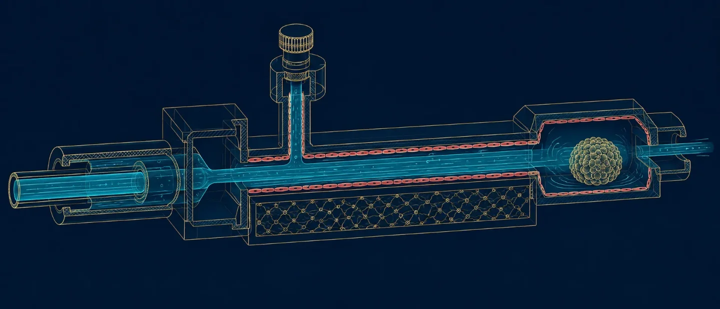
The authors built a Teflon-AF tube-in-tube saturator that transfers oxygen into the liquid phase before the liquid enters a culture or perfusion circuit. The study combines kinetic measurements with computational fluid-dynamics simulations to predict oxygen concentration under changing temperature, flow rate, and tube length. 1
Key finding
The saturator reached near-saturation oxygen concentrations in the liquid phase. The device was most sensitive to changes in temperature, flow rate, and tube length at low oxygen concentrations, while the sensitivity decreased as the liquid approached saturation. The computational model agreed with the kinetic experiments and predicted oxygen concentrations across operating conditions. 1
Why it matters
Oxygen supply can be specified before a liquid reaches an endothelial channel, cell aggregate, or graft compartment. That arrangement lets a vessel-on-chip experiment vary oxygenation independently from channel geometry and flow, instead of treating the oxygen level as an unmeasured consequence of perfusion. The paper also gives a route to connect microscale devices with larger perfusion networks. 1
Main limitation
The preprint validates an oxygen-control device and a predictive model. It does not test an islet, a stem-cell-derived beta-cell graft, a pancreatic endothelial subtype, or a vessel-formation endpoint. The abstract also reports no cell-culture effect size or sample count, so the next experiment still has to establish how the chosen oxygen trajectory changes the biological readout of interest.
Read the original bioRxiv preprint.
2. Hydrostatic pressure changes what low shear means
Evidence class: Adjacent vessel-on-chip mechanobiology
Authors / institution / date: P. Vasanthi Bathrinarayanan, Thomas Abadie, D. Vigolo, M. J. H. Simmons, and L. M. Grover; University of Birmingham and University of Sydney; bioRxiv posting dated 1 September 2026. 2
System / model
The device used a 1,000 μm-wide, 100 μm-high, 17 mm-long microfluidic channel lined with HUVECs. A flow rate of 13 μL/min generated a peak shear stress of 1.4 dyne/cm² in the imaged region. Raising the outlet tube created hydrostatic pressure of about 3,972 Pa at the inlet and 3,924 Pa at the outlet. The authors measured VE-cadherin junctions, YAP1 localisation, EPS8 association, endothelial permeability, and THP-1 monocyte adhesion. 2
Key finding
Adding elevated hydrostatic pressure to low shear produced serrated, finger-like VE-cadherin junctions, more nuclear YAP1, and increased cytoplasmic EPS8-VE-cadherin colocalisation. The pressure condition raised Avidin-FITC permeability by about 2.45-fold relative to the control and increased THP-1 adhesion by about 3.6-fold. Yoda1, a Piezo-1 agonist, reduced permeability to about 1.57-fold relative to control and reduced monocyte adhesion by 2.2-fold relative to the pressure condition. Under low shear without elevated pressure, Yoda1 instead increased permeability by about 2.5-fold and did not significantly increase THP-1 adhesion. Each response came from three experimental repeats, with at least 50 cells analysed per repeat for the junctional measurements. 2
Why it matters
The paper separates shear stress from hydrostatic pressure as two experimental inputs. That distinction matters for islet and beta-cell vascular models because a flow rate alone does not describe the mechanical environment at the endothelial interface. Barrier permeability and immune-cell adhesion can move in different directions when Piezo-1 is activated under different pressure conditions. 2
Main limitation
The model uses HUVEC monolayers and short exposures rather than pancreatic endothelial cells, islets, or transplanted grafts. The perturbations rely on Yoda1 and Wortmannin, so the proposed Piezo-1 and PI3K mechanisms still need genetic or isoform-specific tests. The three-repeat design gives useful direction-of-effect measurements, while the preprint does not establish how the response scales across donors, longer culture periods, or a vascularized islet construct.
Read the original bioRxiv preprint.
3. Matrix patterning selects for endothelial invasion
Evidence class: Adjacent vascular-materials and microfluidic method
Authors / institution / date: Claudia A. Garrido, Daniela S. Garske, B. Haessel, C. Bastard, J. Kamp, Laura De Laporte, Georg N. Duda, Katharina Schmidt-Bleek, and Amaia Cipitria; Charité – Universitätsmedizin Berlin, DWI Leibniz Institute, and Biogipuzkoa; bioRxiv posting dated 3 September 2026. 3
System / model
The authors made patterned interpenetrating networks from covalently crosslinked alginate and physically crosslinked collagen. Norbornene- and tetrazine-functionalised alginate supported two crosslinking routes: degradable links containing matrix-metalloproteinase-sensitive peptides and slower non-degradable links. Photolithography then patterned degradation, collagen fibrillogenesis, microarchitecture, and matrix viscoelasticity inside a microfluidic platform that modelled an early-healing setting. 3
Key finding
Endothelial cells invaded and proliferated in a spatially controlled manner. Regions that combined collagen fibrillogenesis, alginate degradability, and the selected viscoelasticity produced endothelial invasion similar to the pattern reported in vivo after injury. The abstract gives no effect size or sample count. 3
Why it matters
The result keeps matrix degradation, collagen organisation, and viscoelasticity as separate design variables. A vascularized islet construct can therefore test whether endothelial invasion depends on a degradable path, a fibrillar collagen structure, or the time-dependent mechanics of the matrix, rather than reducing all three properties to a single stiffness value. The microfluidic compatibility also makes the material suitable for coupling matrix patterning with controlled flow. 3
Main limitation
The assay models endothelial invasion during an injury-like process. It does not contain islets, stem-cell-derived beta cells, pancreatic support cells, or a demonstrated perfused vessel network. The study is also a bioRxiv preprint and has not been certified by peer review. The paper can define a matrix test matrix; it cannot establish graft vascularization efficacy.
Read the original bioRxiv preprint.
4. Beta cells transfer mitochondria to islet macrophages
Evidence class: Adjacent beta-cell/islet immune and homeostasis biology
Authors / institution / date: Lauar de Brito Monteiro, Anne-Sophie Archambault, Paula Ma, Galina Soukhatcheva, Derek Dai, Jane Velghe, Dorian Izerable, Majid Mojibian, Annette E. Patterson, Ramon I. Klein Geltink, and Cameron Bruce Verchere; University of British Columbia; bioRxiv posting dated 3 September 2026. 4
System / model
The authors used mice with beta-cell-specific mitochondrial GFP expression and complementary in-vitro experiments. The design tracked mitochondrial transfer from beta cells to islet macrophages in vivo and in vitro, then used RNA sequencing to examine macrophages that had received beta-cell-derived mitochondria. 4
Key finding
Beta cells transferred mitochondria to islet macrophages in both settings. Diabetogenic stressors did not change the frequency of transfer. Macrophages containing beta-cell-derived GFP had higher protein-synthesis rates, and RNA sequencing identified increased expression of activity-regulated cytoskeleton-associated protein, or Arc. Disrupting actin-cytoskeleton dynamics prevented the transfer. 4
Why it matters
A graft-cell survival readout can reflect communication with resident or recruited immune cells as well as oxygen delivery and vessel formation. The preprint gives vascularization experiments another reason to measure macrophage state and beta-cell–macrophage contact: a change in insulin output or cell survival may include an immune-homeostasis component that vessel density alone cannot identify. 4
Main limitation
The preprint reports beta-cell–macrophage communication and immune regulation. It reports no endothelial cells, vessel formation, perfusion, transplantation, or stem-cell-derived beta-cell model. The page states that the work has not been certified by peer review and discloses that Cameron Bruce Verchere is a founder, shareholder, and advisory board member of Integrated Nanotherapeutics. 4
Read the original bioRxiv preprint.
What this week changes
The four papers keep four variables apart. The oxygen-saturator paper makes dissolved oxygen an upstream perfusion input. The endothelium-on-chip paper shows that low shear and hydrostatic pressure can produce different barrier and immune-cell responses. The patterned hydrogel paper separates collagen fibrillogenesis, degradation, and viscoelasticity. The macrophage paper adds beta-cell–immune communication as a possible contributor to survival and function.
None of the four papers measures direct revascularization of a transplanted islet or vascularization efficacy for a stem-cell-derived beta-cell or stem-cell-derived islet graft. This week's actionable result is a measurement plan: report oxygen concentration, perfusion continuity, shear stress, hydrostatic pressure, endothelial identity, matrix mechanics, immune-cell state, and graft vessel formation as distinct readouts. A stronger beta-cell survival or insulin signal becomes easier to interpret when those variables are measured separately.
References
- 1
- 2
- 3
- 4
This story was produced automatically by a channel. One sentence is all it takes for Neodrop to keep producing for you.
Related content
More from this channel
- Bifurcation dynamics, endothelial transition, and stem-cell vascular therapy: three new preprints
- Weekly scan: pericyte HGF, human pancreatic interfaces, and the vascularization gap
- Weekly scan: four new vascular-chip and islet papers, with no direct SC-islet vascularization hit
- Weekly scan: VEGF-A priming moves islet reperfusion earlier, while graft niche and printing stay separate problems
- Weekly scan: perfusion, matrix mechanics, and adjacent islet-replacement signals