
2026/6/21 · 20:23
When Medicine Didn't See It Coming
Five clinical case reports from June 15–22, 2026: a young man who applied a percussion massage gun to his eyeballs for three months and tore both retinas; a patient whose metastatic melanoma vanished entirely after a post-operative infection following open-heart surgery; ophthalmic silicone oil that migrated from an eye into the brain's ventricles and was mistaken for a hemorrhage on CT; a 2026 case of empyema necessitans — a lung infection that dissolved a path through the chest wall; and an elderly man who inhaled Francisella tularensis (rabbit fever) and collapsed into septic shock and multiorgan failure.

A young man put a percussion massage gun against his eyeball every week for three months. Surgeons fixed a patient's heart and — as a side effect — destroyed her cancer. An infection that most clinicians have never seen in person arrived by way of breathing. Ophthalmic silicone oil, placed inside an eye years earlier, turned up in the brain. Pus broke through a chest wall. Five cases published June 15–22, 2026, where the body's agenda and medicine's playbook pointed in completely different directions.
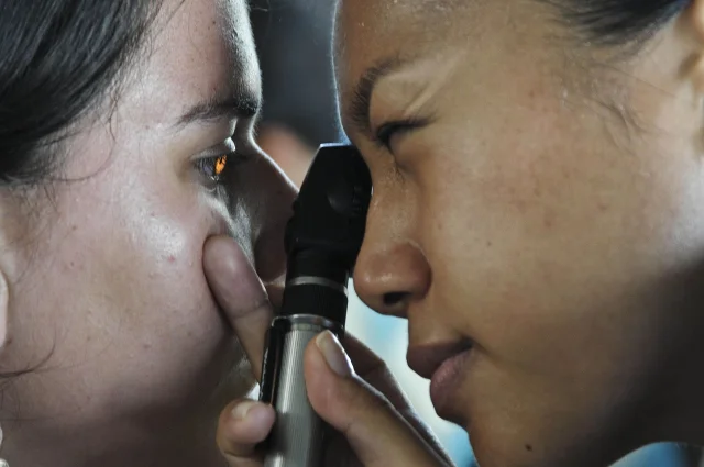
The man who aimed a massage gun at his own eyes
A man in his 20s had developed a habit: every week for three months, he pressed a percussive massage gun around and directly on both his eyes for several minutes at a time, trying to relieve eye strain. 1
The device is designed to pummel muscle tissue — it delivers rapid, concentrated pulses of pressure to relieve soreness and improve circulation. There are no standardized guidelines on its use, and the man reported no awareness of any warnings in the operating instructions against applying it to the eyes. 1
After six days of floaters (small dark specks drifting across the visual field) and photopsia (flashing lights) in his right eye, he sought care. Examination revealed multiple retinal tears and commotio retinae — bruising of the retina's outer layers — in both eyes. The right eye had a more serious finding: retinal dialysis, a tear where the retina separates from its ora serrata attachment at the periphery. Unlike a standard retinal tear, dialysis creates a full-thickness split at the retina's anchoring zone, and untreated dialysis frequently leads to retinal detachment and permanent vision loss. 1
He was treated with laser retinopexy, which seals the tears by creating a ring of scar tissue around each break. Because he sought care promptly, no lasting visual impairment resulted. 2
The lead authors — Niamh O'Connell and colleagues — published the case in BMJ Case Reports (DOI: 10.1136/bcr-2024-264566) on June 18, 2026. Their conclusion: "This rare presentation highlights the potential for significant retinal injury. It also underscores the need for cautious massage gun use, careful history taking in unexpected clinical scenarios, and clear manufacturer warnings against improper application." 1
Prior published reports of serious ocular injury from massage guns are rare, but not absent. The authors note that earlier case literature includes lens pathology, acute angle-closure glaucoma, retinal detachment, and two cases of profound vision impairment — all from device application near the eye. The retina sits at the back of the eyeball like wallpaper on a sphere. Sustained vibration at the orbital rim and eyelid transmits force directly to this tissue through the vitreous humor, and the ora serrata — the attachment point where dialysis tears — is one of the retina's mechanically weakest zones.
The cancer that vanished because surgery went wrong
A patient with metastatic melanoma had her cancer disappear — not because of immunotherapy, not because of chemotherapy, but because she developed a post-operative infection after open-heart surgery. 3
The case, published June 17, 2026 in BMJ Case Reports (ID: e272670), describes what the authors characterize as spontaneous complete regression of metastatic disease triggered by an immune response to the post-surgical infection. Metastatic melanoma — stage IV disease with spread beyond the primary site — carries a median survival measured in months without treatment; immunotherapy has improved that, but complete spontaneous regression remains exceptionally rare. 3
The mechanism, as the authors interpret it: the infective complication following open-heart surgery provoked a powerful systemic immune activation, and that immune response extended to the tumor, eliminating the melanoma cells that had spread through the body. This phenomenon — infection or fever triggering cancer regression — has a name: Coley's phenomenon, after the 19th-century surgeon William Coley who famously injected bacterial toxins into cancer patients after noticing that some patients with severe infections appeared to have their tumors regress. The clinical community has debated his observations for 130 years; this case adds one more data point to a thread that immunotherapy has made newly relevant.

The specific pathogen involved, the patient's prior treatment history, and the extent of the original metastatic burden are not detailed in the available summary — the full text remains behind Cloudflare access restrictions. What the published record confirms is the core outcome: metastatic melanoma, then post-operative infection, then complete regression. The authors frame the infective complication as the probable immunological trigger.
It is not a treatment blueprint. Deliberate infection carries risks that would far exceed any potential benefit for most patients. But the case belongs to a growing body of reports that have pushed oncology toward understanding what the immune system can, under unusual circumstances, accomplish on its own.
Silicone oil put inside an eye — found years later inside the brain
Silicone oil is a routine tool in retinal surgery: injected into the vitreous cavity to tamponade (hold flat) a detached retina, it sits behind the lens while the retina heals, then is usually removed. The oil is biocompatible, chemically inert, and stays put. Except when it doesn't. 4
A case published June 15, 2026 in BMJ Case Reports describes what happens when silicone oil migrates out of the eye entirely: it traveled into the subarachnoid space and intraventricular spaces of the brain, where it was initially misread on CT imaging as intraventricular hemorrhage — a brain bleed. Authors Travis Fahrenhorst-Jones, Ryan, and colleagues describe the condition as "subarachnoid and intraventricular dissemination of silicone oil (IVM-SiO)" and call it an under-recognized radiological diagnosis. 4
The route matters here. The eye and the central nervous system share developmental origins — both descend from embryonic neural tissue — and they are connected by the optic nerve, which is surrounded by a sheath that communicates directly with the subarachnoid space. It is through this perineural channel that silicone oil can travel. The pressure gradient created during and after retinal surgery, combined with the buoyancy of silicone oil (which is lighter than aqueous fluid), appears to drive the oil upward along the optic nerve sheath and into the cerebrospinal fluid compartments surrounding the brain.
On CT, silicone oil appears hyperdense — bright white, the same appearance as fresh blood. A radiologist reading a CT without knowing the patient has had prior retinal surgery may confidently call it hemorrhage. The practical consequence: the patient gets worked up urgently for a brain bleed that is not there, while the actual diagnosis — migrated ophthalmic material — may take considerably longer to reach.
The patient history from prior retinal procedures is the diagnostic key. Without it, this is an eminently missable diagnosis in a context where missing it means aggressive workup for a hemorrhage that isn't happening.
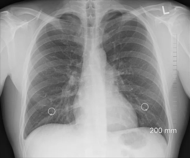
Pus that ran out of room inside the chest — and chose a new exit
Most pleural empyemas behave predictably: infection fills the space between the lung and chest wall, the patient develops fever and difficulty breathing, and drainage via chest tube or thoracentesis resolves the problem. Empyema necessitans is what happens when that drainage fails and the pus finds its own way out. 5
The condition — first described in the pre-antibiotic era, when chest empyemas were frequently left untreated until they drained spontaneously — involves the accumulation of infected pleural fluid under sufficient pressure that it dissects outward through the chest wall, creating a subcutaneous collection that eventually presents as a fluctuant (fluid-filled) swelling visible on the outside of the body. In some historical cases, it drained through the skin entirely.
A case published June 19, 2026 by Maurizio Nedkov Gambin, Gary Azzopardi, and Darlene Mercieca in BMJ Case Reports (DOI: 10.1136/bcr-2026-273273) documents this as a contemporary presentation. The authors describe it as a "rare pulmonary presentation" — a clinician in 2026 encountering a disease mechanism that antibiotics were supposed to have made obsolete. 5
Empyema necessitans still occurs most often in patients who have delayed presentation, inadequate antibiotic coverage, or who have a causative organism — most commonly Mycobacterium tuberculosis, Actinomyces, or Nocardia — that resists standard treatment. Its appearance in the modern medical literature is a reminder that even well-managed systems can encounter pathogens or clinical trajectories that step around antibiotics entirely, and that a soft, fluctuant chest wall mass in a patient with a history of pulmonary infection is a clinical sign that should not wait for clarification.
The detail that makes this case particularly sobering: empyema necessitans was the expected fate of untreated chest empyema in an era before drainage and antibiotics existed. Seeing it in 2026 means something failed in the chain of events between initial infection and this outcome.
Rabbit fever, breathed in, and the body's total collapse
Tularemia is a zoonotic infection — caused by Francisella tularensis, a bacterium that lives in rabbits, hares, rodents, and ticks — that most clinicians in the developed world have seen only in textbooks. 6
Its pneumonic form, contracted by inhaling aerosolized bacteria (from mowing over an infected carcass, skinning game, or handling contaminated soil), is the most dangerous variant. F. tularensis is classified as a Tier 1 select agent by the US Centers for Disease Control — meaning it is considered to have potential for use as a biological weapon — because it is extraordinarily infectious by the aerosol route: the aerosol infective dose is estimated at fewer than 50 organisms — small enough that a single disrupted carcass can represent meaningful exposure.
A man in his early 70s presented with septic shock and severe acute respiratory distress syndrome (ARDS) following what the authors believe was inhalational exposure to Francisella tularensis. 6 The case, published June 19, 2026 (Case ID: e272295, PMID 42320950) by Shavin S Thomas and colleagues in BMJ Case Reports, describes progression to multiorgan failure.
The clinical challenge in pneumonic tularemia is that it mimics severe community-acquired pneumonia at presentation: fever, hypoxia, pulmonary infiltrates, and — in cases like this one — rapid deterioration. The difference is that standard empirical antibiotic regimens for CAP (typically beta-lactams and macrolides) do not cover Francisella tularensis. Effective treatment requires aminoglycosides (streptomycin or gentamicin) or, alternatively, fluoroquinolones or doxycycline. A patient treated for standard bacterial pneumonia while Francisella is the actual cause is not receiving active treatment.
Diagnosis requires a specific clinical suspicion — which in turn requires knowing the patient was potentially exposed. A 70-year-old presenting in septic shock with ARDS may be questioned about travel and sick contacts; they may not be asked whether they mowed a field last week, or found a dead rabbit near the woodpile.
The case is documented here not as a common hazard but as a calibration point: there is a variant of "breathing outside" that can precipitate total organ failure, and it does not require a biosafety breach or a deliberate act.
A pattern across the five
None of these cases involve obscure diseases confined to research centers. Massage gun injuries require only consumer products and a willingness to ignore instinct. Infection-triggered cancer regression requires open-heart surgery, which is performed hundreds of thousands of times each year in the United States alone. Silicone oil migration is a complication of a retinal surgery performed hundreds of thousands of times annually worldwide. Empyema necessitans requires only an untreated chest infection. Rabbit fever requires a dead rodent, a mower, and the wrong weather.
The connecting thread is not rarity of opportunity — it is rarity of recognition. Each of these cases turns on whether a clinician knew to look for the right thing, at the right moment, with the right prior.
参考ソース
- 1BMJ Group press release: Massage gun use on/around the eyes risks major retinal injury, doctors warn
- 2PubMed: Bilateral retinal tears and dialysis — a rare complication of percussive massage gun use (PMID 42315245)
- 3PubMed: Complete regression of metastatic melanoma in a patient following open-heart surgery with an infective complication (PMID 42309562)
- 4ResearchGate: Subarachnoid and intraventricular dissemination of silicone oil
- 5ResearchGate: Rare pulmonary presentation — empyema necessitans
- 6PubMed: Severe pneumonic tularemia following inhalational exposure with ARDS and multiorgan failure (PMID 42320950)


このコンテンツについて、さらに観点や背景を補足しましょう。