2026/7/4 · 0:17
Broca's area: why the brain's language center is not one box
Broca's area began as a landmark lesion finding, but modern MRI and fMRI show that the famous left frontal language region is not one uniform speech-production module. This article follows Broca's patient evidence, the re-examination of his cases, and the current view of neighboring language-selective and domain-general systems.

Broca’s best-known patient could understand more than he could say. He was not globally confused, and he was not unable to move his mouth. Yet when he tried to speak, the same syllable came out again and again: "tan." That clinical mismatch gave Paul Broca a powerful idea in 1861: language could break in a specific way when a specific part of the left frontal lobe was damaged. 1
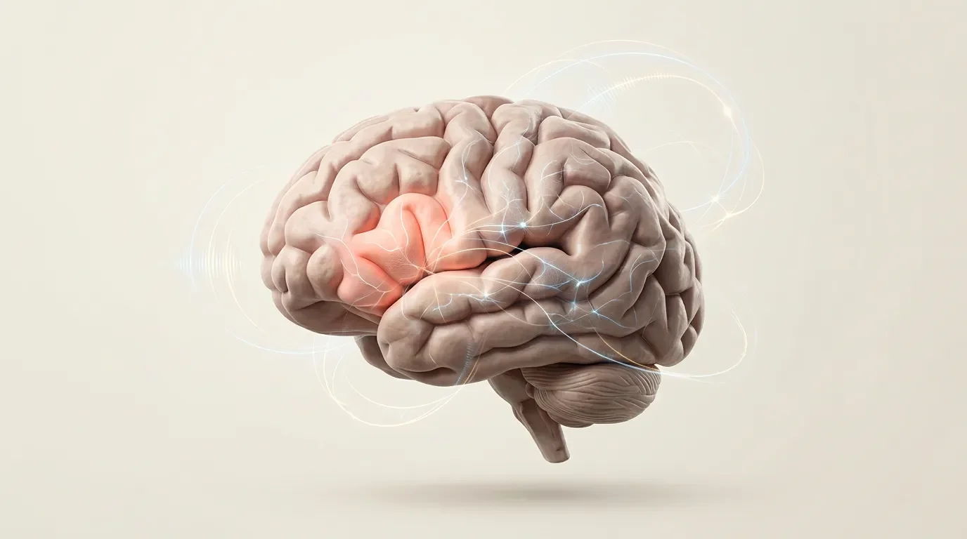
The modern lesson is subtler. Broca’s area matters for language, but it is not a single speech-production box. It is a patch of left inferior frontal cortex where language-selective tissue, task-control tissue, white-matter connections, and person-to-person anatomical variation sit close together. The old localization story was the starting point. The current concept is a warning about how easy it is to turn a useful landmark into an oversimplified module.
What Broca’s area is
Broca’s area usually refers to the posterior part of the left inferior frontal gyrus, especially the pars opercularis and pars triangularis, often mapped approximately to Brodmann areas 44 and 45. In most people it lies above and behind the left eye, in the dominant hemisphere for language. 2
That anatomical definition is useful but incomplete. A gyrus is a fold of cortex; a Brodmann area is a map based on cell structure; a language function is something the brain does. Those three boundaries do not line up perfectly. Calling all of them "Broca’s area" can make a messy region look cleaner than it really is.
In everyday teaching, Broca’s area is often paired with Wernicke’s area: Broca for production, Wernicke for comprehension. That pairing is memorable, but it compresses too much. Speech production needs motor planning, articulation, word retrieval, syntax, auditory feedback, and control over competing responses. Comprehension also recruits frontal language regions when sentences become complex or when the listener must choose among competing interpretations. The region is involved in language, but not in the cartoon sense of a little speech machine.
The landmark study: a lesion that made language localizable
Broca’s 1861 report centered on Louis Victor Leborgne, the patient later nicknamed "Tan." Leborgne had lost fluent speech while apparently preserving much of his understanding. After Leborgne died, Broca examined his brain and reported damage in the left frontal lobe. 1
This mattered because it gave localization a clinical foothold. If a patient can lose articulate speech while other mental abilities remain partly intact, then language is not an indivisible mental faculty spread evenly through the whole brain. Broca’s claim helped shift the debate toward brain-behavior mapping: a particular mental operation could be linked to a particular neural injury.
The evidence was not as simple as the textbook version. Broca initially used the term aphemia for the loss of articulated speech. That is narrower than "all language." The lesion was also not a clean experiment; stroke damage does not respect the tidy borders that later atlases draw. Still, the case made a durable point: fluent speech can fail in a selective, anatomically patterned way.
What modern MRI changed about the old case
In 2007, Nina Dronkers and colleagues used high-resolution MRI to re-examine the preserved brains of Broca’s two historic patients, Leborgne and Lelong. They found that both lesions extended deeper and more medially than Broca could see from the surface. They also reported inconsistencies between the surface area Broca identified and what later researchers often call Broca’s area. 3
That does not make Broca wrong. It changes the interpretation. The patients’ speech problems may have depended not only on cortical tissue in the inferior frontal gyrus, but also on nearby and deeper structures plus disrupted connections. In lesion studies, a damaged region can be the visible tip of a larger network injury.
This point matters for clinical reasoning. If a stroke patient has nonfluent speech, it is tempting to say, "Broca’s area is damaged." Sometimes that is close enough for a bedside shorthand. For science, it is not enough. The deficit may reflect a larger left frontal-insular-subcortical injury, disconnection from temporal language areas, or damage to regions that support speech planning and control.
The fMRI puzzle: language and control sit side by side
Functional MRI complicated the story in a different way. Many tasks activate the left inferior frontal cortex: sentence comprehension, word generation, arithmetic, working memory, response inhibition, and hard attention tasks. One interpretation was that Broca’s area is a general-purpose control system rather than a language-specific region. Another was that classic group-average imaging blurred together neighboring regions that do different jobs.
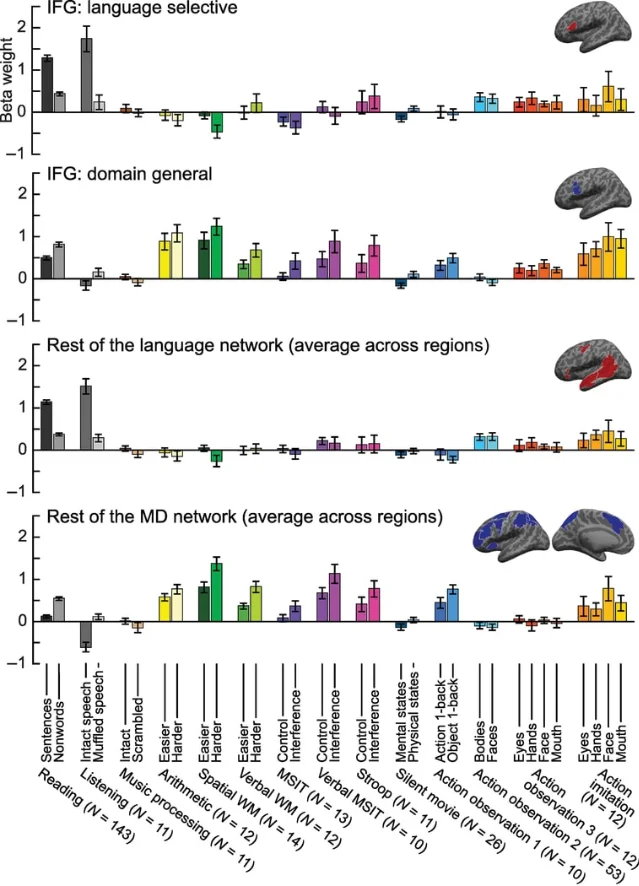
Fedorenko, Behr, and Kanwisher tackled this by defining language regions separately in each participant, rather than assuming that everyone’s language cortex sits in exactly the same atlas location. In their 2011 PNAS study, they used a functional localizer to find each person’s language-responsive regions, then tested whether those regions responded strongly to arithmetic, working memory, cognitive control, and music. They found little or no response in those language regions to the nonlinguistic tasks. 4
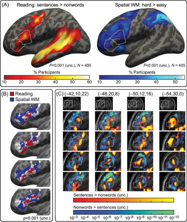
The next step narrowed the question inside Broca’s area itself. In a 2012 Current Biology study, Fedorenko, Duncan, and Kanwisher reported that Broca’s area contains two sets of subregions lying side by side: one selectively engaged by language processing, and another broadly engaged by difficult tasks across domains. 6
This is the central update. The old question was "Is Broca’s area for language or for general cognition?" The answer may be: the label covers nearby pieces of cortex with different profiles. If researchers average across people or use a large anatomical region, those pieces can blur together. If they localize function in each person, the separation becomes easier to see.
What the region may actually be doing
There is no single agreed sentence that captures Broca’s area. Several hypotheses are still alive.
One view emphasizes syntax: the region helps combine words into structured phrases and sentences. Another emphasizes speech production: it supports planning or sequencing the movements and representations needed for fluent utterances. A third emphasizes cognitive control in language: when a sentence is ambiguous, when a speaker must select one word over a competitor, or when grammar conflicts with habit, left inferior frontal cortex may help bias the system toward the right interpretation or response.
The side-by-side model reconciles part of the disagreement. Language-selective frontal regions can participate in the language network, while neighboring multiple-demand regions help when a task is hard, conflict-heavy, or working-memory-intensive. The same experiment can activate both if it uses difficult language material. That does not prove both regions do the same thing.
This is why the method matters. A group-average blob labeled "Broca’s area" can mix language tissue and general-control tissue. A single-subject localizer can ask a cleaner question: which voxels respond more to sentences than to nonwords, and which respond more to general task difficulty? The answer is still imperfect, because fMRI is indirect and slow, but it respects the fact that individual brains do not obey a standard atlas exactly.
Why it matters
Broca’s area is one of the best teaching examples in cognitive neuroscience because it shows both the power and the danger of localization.
The power is real. A specific injury pattern helped reveal that language has neural structure. Broca’s case pushed neuroscience away from vague whole-brain explanations and toward testable brain-behavior relationships.
The danger is also real. Once a label becomes famous, it can hide variation. "Broca’s area activated" does not automatically mean "speech production happened." "Broca’s area damaged" does not automatically explain every symptom in Broca’s aphasia. A better habit is to ask which subregion, which task contrast, which patient lesion, and which network connection are being discussed.
That habit also changes how we read the broader language system. Language is not stored in one cortical address. It depends on a left-lateralized network distributed across frontal and temporal cortex, with nearby control systems that become relevant when language is hard to process or produce. MIT 9.13’s language module frames the topic in exactly this larger way: language in the brain, and the long-standing relationship between thought and language, rather than a single isolated spot. 7
Landmark paper
Broca, P. (1861). "Remarks on the Seat of the Faculty of Articulated Language, Following an Observation of Aphemia." The historical report introduced the lesion-based argument that articulated speech depends especially on the left frontal lobe. 1
Course connection
This concept belongs with MIT 9.13’s language unit. It connects the course’s larger themes: functional specificity, patient evidence, fMRI localization, and the limits of simple one-region explanations. The clean takeaway is not "Broca’s area equals speech." It is that a famous anatomical landmark can contain neighboring systems, and cognitive neuroscience has to separate them with better tasks, better localization, and better lesion interpretation. 7
参考ソース
- 1Remarks on the Seat of the Faculty of Articulated Language
- 2Brain’s language center has multiple roles
- 3Paul Broca’s historic cases: high resolution MR imaging of the brains of Leborgne and Lelong
- 4Functional specificity for high-level linguistic processing in the human brain
- 5Broca’s Area Is Not a Natural Kind
- 6Language-selective and domain-general regions lie side by side within Broca’s area
- 7Lecture 18: Language I
このチャンネルのその他のコンテンツ
関連コンテンツ
- ログインするとコメントできます。