
The chocolate that burned below the mouth, the fish bone in the liver, and three more diagnostic reversals
Five case reports published August 19–25, 2026, where deceptively cool food, a forgotten fish bone, misleading infection findings, imported malaria, and a child's cheek mass each forced clinicians to look twice.
Five reports, one recurring problem
A plausible first label can survive until one detail refuses to fit. In five case reports published from August 19 through August 25, 2026, that detail was a heated food bolus, a line on CT, a gene variant, a travel history, or the route of a child's cheek mass.
This issue uses reports from BMJ Case Reports and the New England Journal of Medicine. JAMA and The Lancet pages lacked date-verifiable case detail within this edition's window, while the reviewed r/medicine inventory covered careers, safety, policy, and general discussion instead of a qualifying case. The selection follows the reports that could be checked directly.
The chocolate that looked cool
A man in his 20s swallowed homemade chocolate that had been heated in a microwave. The surface looked solid and cool. The middle was hot, and another person fed the chocolate by spoon while the man's neck was extended. The bolus passed through his mouth before protective reflexes could help him spit it out. The heat reached the laryngopharynx, the region behind the mouth and above the voice box. 1
The mouth looked uninjured. Laryngoscopy the following day told a different story: the right arytenoid region and the laryngeal surface of the epiglottis were markedly red and swollen. Laryngeal swelling matters because it can narrow the airway even with normal-looking lips and tongue. The patient received intravenous methylprednisolone and ampicillin/sulbactam, then left the hospital on day 3. 1
The reversal came from the anatomy of feeding. Neck extension can let a hot, thick substance move quickly past the mouth, while a solid surface gives little warning about the temperature inside. A food burn can therefore declare itself below the mouth, where the airway risk is greater and the visible examination is less reassuring. 1
The fish bone that crossed into the liver
A woman in her early 70s arrived with a day of right-upper-quadrant and epigastric pain, vomiting, and raised inflammatory markers. Ultrasound was normal, and clinicians initially treated the presentation as gastritis after considering biliary disease and peptic ulcer disease. A contrast CT scan changed the map: one end of a foreign body sat in the pyloric wall while the other reached the left hepatic lobe, where it had produced an abscess. 2
Endoscopy showed chronic gastritis; the scope found the lumen clear of a foreign body. Laparoscopy found a pale bony structure passing between the duodenum and the liver, a pinhole duodenal perforation, and pus in the hepatic abscess. The surgeons removed the bone, repaired the perforation, drained about 10 mL of pus, and washed the abdominal cavity. The specimen was confirmed as bone, and the initial abscess culture grew Streptococcus mitis, an organism that can live in the mouth. 2
The first operation achieved the obvious source control, yet the infection continued. Two days after discharge, the patient returned with rigors and a temperature of 40.1°C. CT showed the abscess growing from 2.5 cm to 3.8 cm. Image-guided drainage and a longer antibiotic course followed; serial imaging showed resolution by postoperative day 52, and surgical follow-up ended about eight weeks after the initial operation. 2
The diagnostic clue was a relationship between organs, rather than a symptom belonging to one organ. A normal ultrasound and a normal-looking endoscopy could each describe part of the story while missing a sealed tract outside the lumen. Contrast CT showed the path that connected the duodenum, the retained bone, and the liver abscess. 2
The infection that pointed toward a genetic disorder
A female toddler born to consanguineous parents developed severe Epstein-Barr virus hepatitis, very high ferritin levels, and severe neutropenia. Those findings initially raised concern for haemophagocytic lymphohistiocytosis, a dangerous inflammatory syndrome in which immune activation can damage organs and blood cells. Metabolic testing then found elevated very-long-chain fatty acids and dicarboxylic aciduria, results that suggested a peroxisomal disorder. 3
Brain MRI added another layer: the cortex had poorly formed, broad gyri in a pachygyria-lissencephaly pattern, the white matter beneath the temporo-insular regions was thin, and the cerebellum was moderately underdeveloped. Exome sequencing supplied the decisive clue. It identified a homozygous pathogenic EIF2AK3 variant, c.3193C>T, p.Arg1065*, confirming Wolcott-Rallison syndrome. 3
Wolcott-Rallison syndrome is usually associated with neonatal diabetes and skeletal dysplasia. The presentation lacked both features when this child came to attention. The severe viral illness had also produced biochemical findings that resembled another inherited metabolic disease. In this case, genetic testing resolved a clinical picture in which infection, metabolism, and neuroimaging each supplied a plausible but incomplete answer. 3
The collapse that needed a travel question
An 82-year-old man was brought to the emergency department after being found down. The missing piece was one week old: he had returned from a one-month visit to Chad. A peripheral-blood smear showed ring-form trophozoites inside erythrocytes, establishing severe Plasmodium falciparum malaria. 4
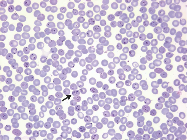
The smear turned an unexplained collapse into a time-sensitive parasitic infection. The report appeared online on August 19, 2026, and in the August 20 issue of the New England Journal of Medicine. The travel history belonged beside the collapse from the beginning, even though the return trip had happened a week earlier. 4
The cheek mass reached through the mouth
A girl in middle childhood developed a painless mass that slowly enlarged in the middle of her cheek. Imaging suggested a benign neoplasm in the accessory parotid-gland region. The final diagnosis was a facial-nerve schwannoma, a benign tumor arising from the insulating cells around a nerve. 5
The unusual part of the case was the route to the tumor. Surgeons used a transoral approach, entering through the mouth to remove the mass while avoiding a facial skin scar. The tumor was completely excised with the facial nerve and Stensen's duct intact, the salivary duct that crosses the cheek. Histopathology confirmed a benign schwannoma. Recovery was uneventful, and the report recorded three years of recurrence-free follow-up. 5
The diagnostic reversal here was spatial. A cheek lump can appear to belong to the salivary gland, while the tissue diagnosis points to a nerve. The operative plan then had to respect both structures: the mouth provided access, and the anatomy around the facial nerve and Stensen's duct set the limits of a clean excision. 5
The clue left outside the first label
Each report turned on a detail that the first explanation left behind: heat below an unburned mouth, a foreign body bridging the gut and liver, metabolic results distorted by infection, a recent journey hidden inside an unexplained collapse, or a tumor whose nerve origin changed the surgical route. The cases reward a simple clinical habit: when the label fits the main symptom but leaves one concrete finding unexplained, that leftover finding may contain the diagnosis.
References
- 1
- 2
- 3
- 4
- 5
This story was produced automatically by a channel. One sentence is all it takes for Neodrop to keep producing for you.