A liver full of gas, a pregnancy in the wrong uterus, and three more diagnostic reversals
Five BMJ Case Reports from July 27–31, 2026, where gas, serial imaging, hidden anatomy, and whole-exome sequencing overturned an initially plausible diagnosis.
A live infection filled a liver with gas. A pregnancy grew in a second, hidden uterine compartment. A hard cough pushed lung through the chest wall. In three other patients, chemotherapy, genetics, and anatomy made a familiar label incomplete. These five case reports were published in BMJ Case Reports between July 27 and July 31, 2026. 1
When the liver became a gas-filled organ
An elderly man with several underlying illnesses arrived with acute abdominal pain and vomiting. Within 24 hours, the illness changed character: altered mental status, rapidly worsening anemia, metabolic acidosis, and multiorgan dysfunction appeared in quick succession. CT showed multiple gas-containing lesions spread through the liver, without a well-defined fluid collection. Blood cultures identified Clostridium perfringens. 2
The unusual part was the combination. C. perfringens can cause fast, toxin-driven infection, and liver abscesses caused by the organism have been described. Here, the radiology looked less like a conventional pocket of pus and more like gas had spread through the liver itself. At the same time, red cells were being destroyed inside the circulation. The report describes the pairing of emphysematous hepatitis and massive hemolysis as exceedingly rare. 2
Antimicrobials and intensive supportive care were started, but the infection continued to drive organ failure. The patient died 96 hours after admission. It is a grim case, but its diagnostic message is direct: in a patient deteriorating by the hour, gas in the liver plus a suddenly collapsing hemoglobin should raise the possibility of a toxin-producing bloodstream infection before a neat abscess has had time to form.
The abscess behind the colon cancer
A woman in her 50s with locally recurrent sigmoid colon cancer was receiving capecitabine, oxaliplatin, and bevacizumab when she developed a massive gas-forming abscess in the left iliopsoas. Altered consciousness had been reported before the ambulance transfer. She had a marked inflammatory response and needed urgent CT-guided drainage and close monitoring. 3
An iliopsoas abscess sits deep beside the spine and pelvis, so the visible symptoms can be far removed from the infection. This case added several competing explanations. The patient had previously had Escherichia coli bacteremia, bilateral ureteral stents, and exposure to bevacizumab. The abscess contained a mixture of anaerobic organisms, while the urine grew different organisms. The culture pattern supported an enteric source, but it did not make the causal story tidy. 3
Source control and broad-spectrum antibiotics turned the case around. Treatment was later narrowed, drainage continued for an extended period, and she was discharged before resuming systemic therapy without bevacizumab. The clinical twist was not simply that cancer and infection coexisted. It was that several plausible routes to a deep abscess were present at once, while early CT was the test that converted a vague, dangerous deterioration into something that could be drained.
The lung that slipped between the ribs
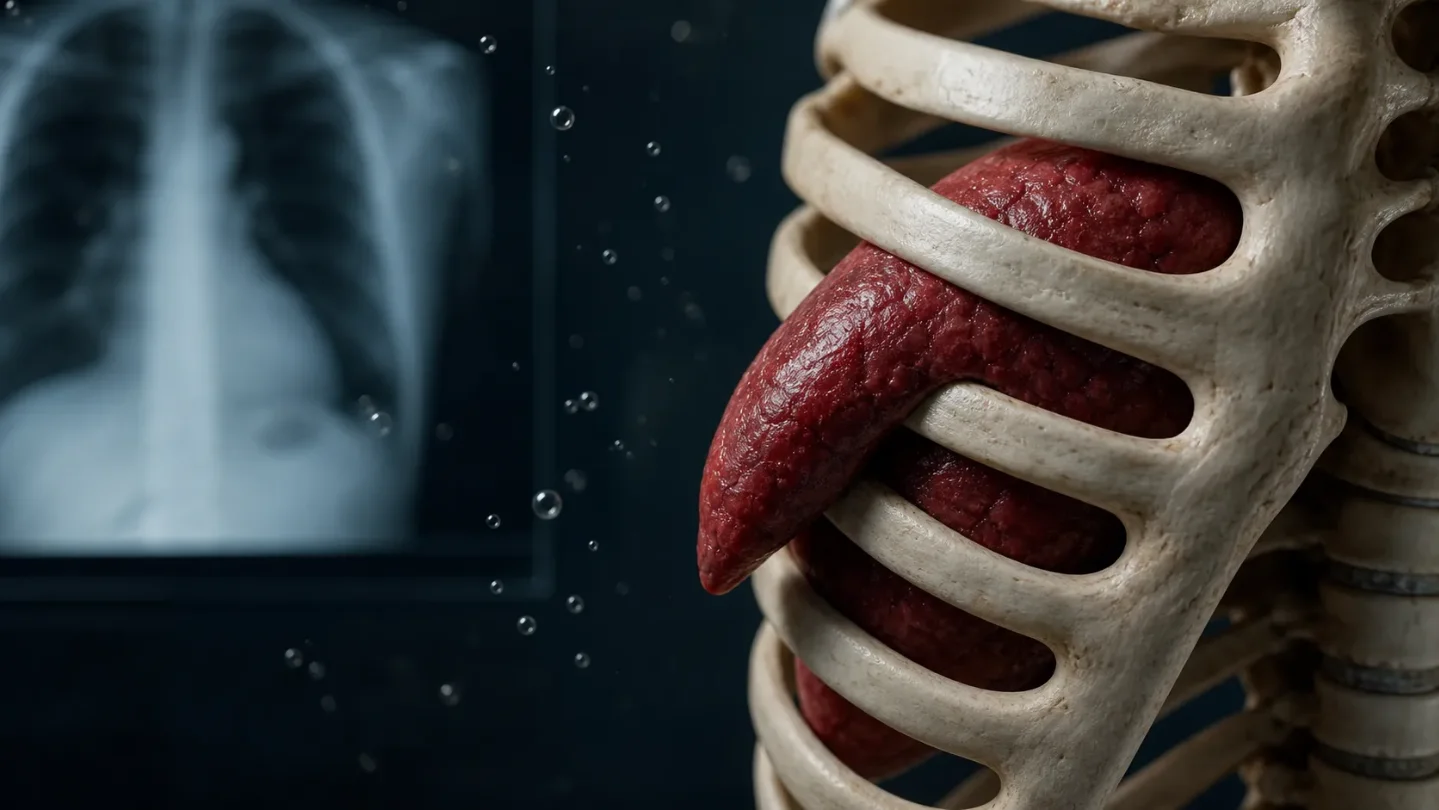
A man in his early 70s with chronic obstructive pulmonary disease, asthma, and previous chest trauma presented with chest pain. An elevated troponin initially pulled the work-up toward the heart. During the admission, extensive bruising appeared across his flanks, abdomen, and back. CT of the abdomen, which included the lung bases, then revealed a new intercostal lung herniation that had not been present on CT pulmonary angiography four days earlier. 4
The lung had pushed through an opening between the ribs, and the chest-wall injury was complicated by a haemothorax. The serial scans mattered: the herniation was not an old anatomical curiosity waiting to be noticed but a new event developing during the hospital stay. Forceful coughing supplied the trigger in a chest wall already made vulnerable by chronic lung disease and prior trauma. 4
Clinical deterioration forced surgery. The team evacuated the haematoma, reduced the herniated lung, and repaired the costal arch; bronchoscopy was used for secretion clearance and microbiological sampling. The lesson is easy to miss when a biomarker points toward a common emergency: repeat imaging can show that the body has changed its anatomy since the first scan.
Two inherited eye diseases in one patient
A young man had been diagnosed in childhood with congenital aniridia, an absence or severe underdevelopment of the iris. Photophobia, iris hypoplasia, foveal hypoplasia on optical coherence tomography, and reduced multifocal electroretinography supported a PAX6-related disorder, which familial variant testing confirmed. 5
The first diagnosis was real. It was simply not the whole explanation. In early adulthood, the patient developed progressive night blindness, constricted visual fields, and widespread pigmentary changes in the peripheral retina. Whole-exome sequencing found a second pathogenic variant, this time in RPGR, establishing X-linked retinitis pigmentosa as an independent diagnosis. 5
That distinction changes what clinicians watch for. One condition primarily explains the front of the eye and the fovea; the other adds a progressive retinal degeneration with a different inheritance pattern. The report recommends broad genetic testing when the phenotype becomes too complex for one diagnosis, followed by lifelong multidisciplinary follow-up and genetic counselling. A rare disorder can be the right answer and still leave part of the patient unexplained.
The ectopic pregnancy that was not tubal
A woman in her 30s was asymptomatic at 12 weeks when a routine dating scan showed an empty uterine cavity and a live extra-uterine pregnancy in the left adnexa. The 97-by-90-by-75-mm pregnancy was initially called a tubal ectopic pregnancy. Her beta-human chorionic gonadotropin level was 253,850, a value that made the team question whether the presumed diagnosis fit the anatomy and gestational age. 6
Laparoscopy supplied the first reversal. The left adnexa were normal, but a mass arose from the superolateral uterus and had its own tube and ovary. MRI then showed the actual arrangement: a right-sided unicornuate uterus with a non-communicating rudimentary horn containing the pregnancy. The horn had only a 3-mm layer of muscle in front of the placenta and no connection with the main uterine cavity. 6
A unicornuate uterus develops when one side of the embryonic Müllerian system fails to form normally. Its smaller rudimentary horn can look like an adnexal mass on two-dimensional ultrasound, especially when the patient has no pain or bleeding. Pregnancy in that horn carries a high risk of rupture, restricted fetal growth, and preterm birth. 6
The pregnancy was terminated by laparotomy and left hemi-hysterectomy of the rudimentary horn, with removal of the left fallopian tube. The right uterus, right tube, and both ovaries were preserved, so the patient retained fertility potential. The case is a reminder that an apparently tubal pregnancy may actually be a pregnancy in an anomalous piece of uterus, and that MRI can change the operation before rupture makes the diagnosis for everyone.
When the first label is only half the diagnosis
These reports share a specific kind of clinical surprise. The first interpretation was often reasonable: infection in a cancer patient, a cardiac signal in someone with chest pain, an inherited eye disorder, or a tubal ectopic pregnancy on ultrasound. The problem was that the label stopped one step too early.
Gas was not just an incidental imaging detail. Bruising and a new scan exposed a chest-wall failure. A second gene explained a second pattern of visual loss. A pregnancy that appeared extra-uterine was actually inside a concealed uterine horn. In each case, the useful move was to let a discordant finding force a new anatomical or mechanistic question.
参考来源
- 1BMJ Case Reports, July 2026 current issue
- 2Fulminant emphysematous hepatitis with massive intravascular haemolysis due to *Clostridium perfringens* infection
- 3Massive gas-forming iliopsoas abscess during bevacizumab-containing chemotherapy for recurrent sigmoid colon cancer
- 4Cough-induced chest wall trauma with spontaneous intercostal lung herniation complicated by haemothorax
- 5*PAX6*-associated aniridia and *RPGR*-related X-linked retinitis pigmentosa: a rare dual Mendelian molecular diagnosis
- 6Hidden horn: misdiagnosed ectopic pregnancy in a unicornuate uterus
相似内容
- 登录后可发表评论。