Six Cases Where Medicine Had No Playbook

Six Cases Where Medicine Had No Playbook

Six clinical case reports from May 17–25, 2026: a lung cancer with an ultra-rare RET mutation that spread across the placenta in the first reported case of its kind; a patient whose esophagus turned coal-black on endoscopy; a right ventricular cardiac tumor that hid behind kidney failure and anaemia; a retina flooded in the "blood and thunder" pattern of central retinal-vein occlusion; a 20-year-old whose routine hip scope ended five years later in amputation for refractory CRPS; and a patient allergic, on paper, to air.

Weirdest Medical Cases
May 25, 2026 · 9:26 AM
4 subscriptions · 2 items
Medicine leaves a long tail. The cases that make it into weekly journals aren't the representative ones — they're the ones where something went fundamentally sideways: a lung tumor that crossed the placenta, a heart tumor that disguised itself as failing kidneys, a retina that flooded with blood in every direction at once. This week's six cases, published or surfaced between May 17 and 25, 2026, span NEJM, BMJ Case Reports, and r/medicine.

Lung cancer crossed the placenta — a first in the medical literature

A pregnant woman was diagnosed with what appeared to be non-small cell lung cancer (NSCLC). After further workup, the histopathology told a different story: the tumor was actually a neuroendocrine carcinoma of the lung — a rarer and often more aggressive subtype — and it carried a mutation in the RET gene, specifically the RET p.M918T variant. 1
Loading link preview…
That mutation, p.M918T, is well-known in oncology — but almost entirely in the context of medullary thyroid carcinoma (MTC), a cancer of the thyroid's calcitonin-producing cells. Finding it as the driver of a primary lung neuroendocrine tumor was already unusual. What came next pushed the case into territory the medical literature had never mapped.
The cancer had spread to the placenta.
Placental metastasis from a maternal malignancy is itself a vanishingly rare event — documented in scattered case reports over decades, occurring most often with melanoma, breast cancer, and leukemia. But a RET p.M918T-mutated neuroendocrine lung tumor with transplacental spread had never been reported. The authors of the BMJ Case Reports paper, published in the May 2026 issue, are explicit: this is the first reported case of this combination in the medical literature. 1
Three independent rarities — driver mutation, tumor type, metastatic route — landed in one patient during pregnancy, where diagnostic latitude is already constrained by fetal considerations. The initial misclassification as NSCLC (a far more common lung cancer category) illustrates the diagnostic challenge: without molecular profiling, a rare variant of a rare tumor looks like the common thing on histopathology slides.

The esophagus that turned black

An endoscope going down a patient's esophagus should find pink, glistening mucosa. What it found instead, in a case published May 23, 2026 in NEJM Images in Clinical Medicine, was a coal-black tube with ulcerated longitudinal necrosis running its length. 2
Loading link preview…
The diagnosis: acute esophageal necrosis (AEN), also called Gurvits syndrome or, bluntly, "black esophagus." AEN occurs when ischemic injury — typically from a combination of compromised blood flow, low systemic perfusion, and mucosal barrier breakdown — attacks the distal esophagus preferentially. The distal esophagus has a relatively sparse blood supply even at baseline, which is why it fails first when perfusion drops.
Authors K. Nie and X. Wang described the endoscopic finding in a case severe enough to warrant publication in the NEJM's Images section: esophagogastroduodenoscopy (EGD, the scope that examines the esophagus, stomach, and duodenum together) revealing diffuse black coloration with longitudinal strips of necrotic, ulcerated tissue. The image is the diagnosis — there is no differential for a fully black esophagus. 2
AEN is rare enough that many gastroenterologists encounter it only once or twice across a career. When it does appear, the patient is almost always severely ill from another underlying cause — sepsis, cardiogenic shock, diabetic ketoacidosis — that has driven the perfusion failure. The black esophagus is the visible index of how sick the rest of the patient is.

The heart tumor hiding behind kidney failure

A 53-year-old man presented with severe anaemia and acute kidney injury. Neither of those findings points at the heart. The workup that followed, however, found a large right ventricular myxoma — a primary cardiac tumor — as the underlying cause of both. 3
Loading link preview…
The case, published in BMJ Case Reports (Vol. 19, Issue 5, 2026), layers two levels of rarity. First: cardiac myxomas are the most common primary tumors of the heart, but the overwhelming majority — roughly 75% — arise in the left atrium. A right ventricular origin already puts this case in a distinct minority. Second: even left atrial myxomas classically present with cardiac symptoms (dyspnea, palpitations, embolic events from fragments breaking off the tumor). This patient's heart tumor generated no cardiac symptoms at all. His presentation was the haematological and renal fallout from a mass the cardiologists hadn't been asked to look for yet. 3
The mechanism linking a cardiac tumor to anaemia and kidney injury involves the tumor's mechanical obstruction of blood flow through the right ventricle and pulmonary outflow tract, causing turbulence that damages red blood cells (a microangiopathic haemolytic pattern) and reduces renal perfusion. The causal chain runs: tumor → turbulent shear stress → haemolysis + hypoperfusion → anaemia + renal failure. Each step is logical in retrospect; none of it announces "cardiac tumor" at the front door.

The retina that looked like a thunderstorm

A healthy fundus — the back wall of the eye as seen through an ophthalmoscope — shows a clean orange-pink surface with a few branching vessels converging on the optic disc. What clinicians call the "blood and thunder" fundus looks nothing like that. In central retinal-vein occlusion (CRVO), an acute thrombus blocks the central retinal vein at or just behind the optic disc, and with no exit route the backed-up venous blood erupts outward across every quadrant of the retina. The result is a fundoscopic image of dense, flame-shaped hemorrhages radiating from the disc in all directions — diffuse, dramatic, and recognizable in an instant. 4
Loading link preview…
The case, published May 14, 2026 in NEJM Images in Clinical Medicine by G. Calvão Santos, illustrates why ophthalmologists reach for the "blood and thunder" descriptor: there is no other fundoscopic finding that fills the retina with hemorrhage this completely or this symmetrically. CRVO is not rare in absolute terms — it is the second most common retinal vascular disorder after diabetic retinopathy — but the complete, florid presentation of a central vein occlusion captured in a published image carries a different weight than the partial or resolving variants most clinicians see. 4
The mechanism is a thrombotic blockade of the point where all retinal venous drainage converges. Once that single exit is occluded, hydrostatic pressure forces blood through capillary walls across the entire retinal surface simultaneously. Vision loss is typically sudden and can range from mild blurring to near-total loss of central vision depending on whether the macula is involved. The clinical urgency is partly about the eye itself — but also about what CRVO signals systemically: hypertension, hyperviscosity states, glaucoma, and hypercoagulable conditions are all implicated, and a new diagnosis of CRVO often prompts a cardiovascular and haematological workup that turns up the underlying driver.

Hip scope, five years of agony, then amputation at 20

A 20-year-old woman underwent hip arthroscopy — a minimally invasive procedure, typically performed to address labral tears or femoroacetabular impingement. What followed was Complex Regional Pain Syndrome Type 1 (CRPS-1): a poorly understood neurological pain disorder characterized by disproportionate, burning pain, skin color and temperature changes, and motor dysfunction in the affected limb.
Her CRPS-1 was refractory. Over five years, she exhausted medical and interventional options — a course that, for severe CRPS, can span sympathetic nerve blocks, spinal cord stimulation, ketamine infusions, and intensive physical therapy. None produced lasting relief. As a final option, she underwent TiTON surgery (Titanium Transcutaneous Osseointegration Nail) followed by leg amputation. The case was documented by Dr. Jason Hoellwarth, MD, an orthopaedic surgeon at Hospital for Special Surgery (HSS), published on the Orthobullets platform, and subsequently discussed on r/medicine (510 upvotes). 5
The r/medicine thread surfaces a clinical tension that the case encapsulates well. Amputation for CRPS is documented and practiced in Europe with some regularity; in the United States, it remains rare enough that many pain physicians consider it outside standard practice. One commenter characterized the underlying pathophysiology: "It's like Charcot foot. A weird neurovasculomuscular problem that is irreparable." Another noted: "In fact, amputations can be a worthwhile treatment for CRPS and they are done in Europe often enough, but not here." 5
The starting point — elective hip arthroscopy in a 20-year-old — is itself worth noting. Hip arthroscopy at that age is uncommon enough that even the r/medicine community flagged it: "Very unusual to need or recommend a hip arthroplasty at 20 yrs of age." CRPS is a known, rare complication of hip arthroscopy, with the literature citing incidence below 1% in most series. That the single worst trajectory from a rare complication of an already-unusual procedure played out fully is what gets a case onto Orthobullets.

One patient, 27 allergies, including air

On May 23, 2026, a physician posted to r/medicine under the title "I think I may have found a new winner for the Most Ridiculous Allergy award." The body of the post: "Amongst her 27 allergies — Air. No, I'm not kidding. Allergy to Air." 6
The post, by user u/SpawnofATStill, collected 510 upvotes. It needs no clinical context. A documented allergy to the atmosphere means the patient's medical record contains a contraindication that cannot be accommodated under any circumstances, anywhere, by any intervention. The practical implication for anyone who encounters this in an EHR — assuming the field is real and not a data entry artifact — is that the allergy list has become a clinical noise floor rather than a clinical tool.

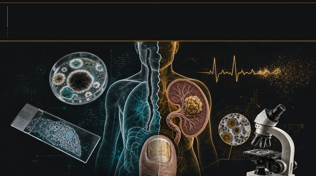
Cover image: AI-generated editorial composite, created for this article.

Add more perspectives or context around this Drop.

  • Sign in to comment.