24/6/2026 · 0:17
Receptive fields: the neuron's window on the world
Receptive fields explain how single neurons answer for specific parts of sensory space. This article traces the idea from Kuffler's retinal center-surround fields to Hubel and Wiesel's V1 edge-selective cells, then shows why context and feedback complicate the clean textbook diagram.

The idea in one sentence
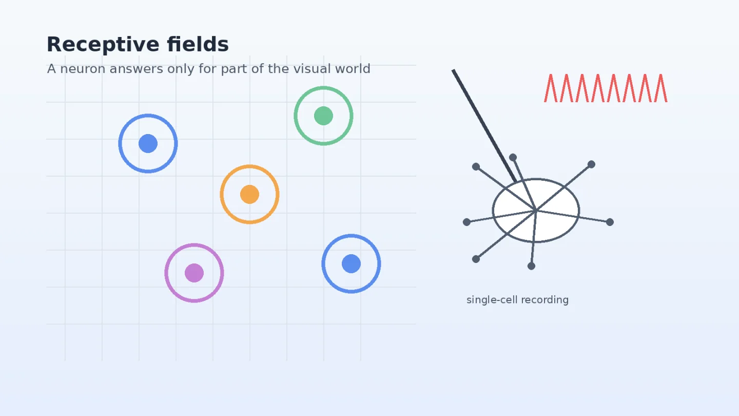
A receptive field is the part of the sensory world where a stimulus can change a neuron's firing. For a visual neuron, that means a particular patch of visual space, plus a rule for what kind of light pattern in that patch matters.
That sounds small, almost like a vocabulary word. It is not. Receptive fields are one of the main reasons neuroscience can connect a physical stimulus, a single cell, and a perception. They let researchers ask a precise question: if I change this little piece of the world, does this neuron care?
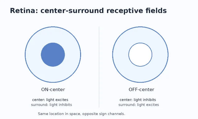
The concept began in the retina. Stephen Kuffler's 1953 cat-retina work described ganglion cells whose responses depended on stimulation of specific retinal regions, and the PubMed record identifies the paper as Discharge patterns and functional organization of mammalian retina in Journal of Neurophysiology.1 A later NCBI textbook chapter summarizes Kuffler's result this way: each ganglion cell responded to a small circular patch of retina, and that patch defined the cell's receptive field.2
What a receptive field is, and what it is not
A receptive field is not a tiny screen painted on a neuron. It is a measurement: present stimuli in different places, record the neuron's spikes, and map where the response changes.
For a retinal ganglion cell, the early answer was often circular. Some cells fired more when light hit the center of the field and less when light hit the surround. Others showed the opposite sign: they were suppressed by light in the center and excited by light in the surround.2 This organization means the retina is not just measuring brightness. It is already comparing center against surround, which makes the system sensitive to contrast and edges.
That is the first lesson: sensory neurons usually do not report raw physical energy. They report structured differences in the world.
The second lesson came from primary visual cortex, or V1. Hubel and Wiesel's 1959 paper, Receptive fields of single neurones in the cat's striate cortex, recorded from cat striate cortex and is listed in PMC with the DOI 10.1113/jphysiol.1959.sp006308.3 They found that many cortical neurons did not respond best to round spots. They responded to bars, slits, or edges in particular positions and orientations.
So the receptive field had become more than a location. It was a location plus a feature preference.
The landmark move: from spots to edges
Hubel and Wiesel's famous trick was not a trick at all. It was patient mapping. They moved light patterns across the visual field while listening to spikes from single cortical neurons. When a spot failed to excite a cell reliably, they tried other shapes. A thin bar at the right angle could make the cell fire vigorously.
Their 1959 paper reported cells in cat striate cortex whose receptive fields had elongated excitatory and inhibitory regions.3 In the 1962 follow-up, Hubel and Wiesel introduced the now-standard distinction between simple and complex cells, while also examining binocular interaction and cortical architecture.4
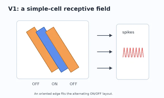
The point was not merely that V1 contains edge detectors. The deeper idea was hierarchical transformation. Retinal and thalamic neurons have center-surround fields. A V1 simple cell can be understood, roughly, as combining inputs arranged in a line. Put enough center-surround inputs side by side, and an oriented edge becomes the best stimulus.
This is why receptive fields became a bridge between biology and computation. They gave neuroscientists a way to talk about the code a neuron appears to implement. They also gave engineers a vocabulary that later reappeared in artificial vision systems, where early layers often learn local filters for edges and texture.
Why receptive fields matter for cognition
Receptive fields help explain how the brain avoids treating vision as a photograph. The eye receives an image, but the brain does not passively copy it inward. At each stage, neurons ask more selective questions.
A retinal ganglion cell may ask, "Is there a light-dark contrast here?" A V1 simple cell may ask, "Is there an edge at this orientation here?" A higher visual-area neuron may ask a broader question over a larger region of the visual field. The field usually gets bigger as information moves up the visual hierarchy, while the preferred stimulus can become more complex.
This matters because cognition needs stable objects, places, faces, words, and actions. Receptive fields are one of the first steps in turning a pattern of light into features that later systems can use. Before the brain can recognize a face, it has to build visual descriptions that are tolerant enough to handle small changes but specific enough not to confuse everything with everything else.
There is a danger here, though. A receptive field is often drawn as a neat patch, but the real response of a neuron depends on the whole experimental situation: contrast, attention, adaptation, eye movements, task demands, and signals arriving from other brain areas. The map is useful because it simplifies. It misleads when we forget that it is a simplification.
The modern complication: the field has a surround
The classic receptive field is the region where a stimulus can directly drive a neuron under a particular measurement procedure. But V1 neurons can also be influenced by stimuli outside that classical region. This is called the extra-classical surround.
Angelucci and Bressloff's 2006 review states that V1 neurons respond best to oriented stimuli of optimal size inside the receptive-field center, but their responses are often suppressed by iso-oriented stimuli in the surrounding region.5 The same review argues that feedforward inputs, lateral V1 connections, and feedback from extrastriate cortex may all contribute to center and surround effects.5
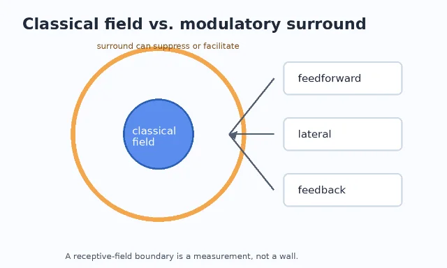
This is where the concept becomes scientifically interesting again. If a stimulus outside the classical field changes the response, then the neuron is not just encoding a local patch. It is also being modulated by context.
That helps explain everyday perception. A gray patch looks different depending on the gray around it. An edge can pop out or disappear depending on the surrounding texture. A line in a natural scene is interpreted differently when it belongs to a tree branch, a shadow, or a face. The early visual system is local, but not isolated.
What scientists still debate
The receptive-field idea is solid. The debates are about mechanism and interpretation.
First, how much of a V1 receptive field is built by feedforward wiring from the lateral geniculate nucleus, and how much is shaped by intracortical computation? Hubel and Wiesel's work made the feedforward story attractive: line up inputs, get orientation selectivity.4 Later work showed that recurrent cortical circuits, inhibition, contrast, and feedback also matter.
Second, should we treat the receptive field as a fixed property of a cell? In a textbook diagram, it looks fixed. In experiments, it can shift with contrast, stimulus history, attention, and task context. The same neuron can look more local or more context-sensitive depending on how it is tested.
Third, how far can the receptive-field idea travel beyond early vision? Researchers use related concepts in audition, touch, spatial cognition, and even deep learning. That broad use is powerful, but it can blur differences. A retinal receptive field measured with spots of light is not the same kind of object as a high-level model unit that responds to complex images.
The safest definition is still the experimental one: a receptive field is the part of stimulus space where changes alter a neuron's response, measured under specified conditions.
Landmark paper
Hubel, D. H., & Wiesel, T. N. (1959). Receptive fields of single neurones in the cat's striate cortex. Journal of Physiology, 148(3), 574-591. The paper's PMC record lists the DOI as 10.1113/jphysiol.1959.sp006308 and the PMID as 14403679.3
Course connection
MIT 9.13, The Human Brain, is built around how perceptual and cognitive abilities are implemented in the brain; the OCW course description says it covers representations, development, and functional specificity across mind and brain.6 Receptive fields are an early visual-system version of that theme: they show how a neuron can represent a specific part of the world and a specific kind of information about it.
Fuentes de referencia
- 1Discharge patterns and functional organization of mammalian retina - PubMed
- 2Retinal Circuits for Detecting Differences in Luminance - Neuroscience - NCBI Bookshelf
- 3Receptive fields of single neurones in the cat's striate cortex - PMC
- 4Receptive fields, binocular interaction and functional architecture in the cat's visual cortex - PMC
- 5Contribution of feedforward, lateral and feedback connections to the classical receptive field center and extra-classical receptive field surround of primate V1 neurons - PubMed
- 6The Human Brain
Añade más opiniones o contexto en torno a este contenido.