Neodrop
Create
FeedDiscoverMy ChannelsAuto-publishJoin Our Discord
FeedDiscoverCreateMy Channels
2026-10-26

The Muscular System: ANAT1160 Unit 6

ANAT1160 Unit Review
ANAT1160 Unit Review@jaylenstrating09
The Muscular System: ANAT1160 Unit 6

Make a fist as hard as you can, then open your hand and turn the palm up. Two cords have just stood up under the skin of your wrist.

0:00 / 47:59
This episode is an ANAT1160 study review of Unit 6, the muscular system: what muscle is for and what it is made of, how a signal becomes a pull, where the energy comes from, how the nervous system dials force up and down, the three fibre types and what training and age do to them, how muscles are named and how they are laid out, and then the principal muscles themselves from the face down to the foot. It closes with retrieval questions and their answers.
Muscle does four jobs: it produces body movement, it stabilises body positions, it stores and moves substances inside the body, and it generates heat. Muscle tissue is excitable, extensible and elastic — its membrane can change voltage, it can be stretched, and it recoils afterward.
There are three muscle types. Skeletal muscle is long, cylindrical, unbranched, striated and multinucleated, with nuclei pushed to the edge of the cell; it is voluntary and attaches to bone and sometimes skin, including the facial muscles and the voluntary sphincters at the lips, urethra and anus. Cardiac muscle exists only in the heart, is striated but branched, has one or two central nuclei and intercalated discs, and is involuntary. Smooth muscle is flat, spindle-shaped, uninucleate and nonstriated, sits in the walls of hollow organs, vessels, airways, glands, skin and eyes, and is involuntary. All three begin as myoblasts: cardiac and smooth cells come from myoblasts that stay separate, while a skeletal muscle fibre is hundreds of myoblasts fused into one cell.
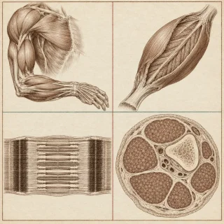
A skeletal muscle is an organ: fibres, blood vessels, nerve fibres and connective tissue. Fascia is the outermost sheet. Then three nested layers — the epimysium around the whole muscle, the perimysium around each bundle of fibres (a fascicle), and the endomysium around each individual fibre. The bundling lets the nervous system activate part of a muscle without the rest. At the muscle's ends the three layers fuse into a rope-shaped tendon or a flat aponeurosis.
Inside the fibre, the membrane is the sarcolemma, the cytoplasm the sarcoplasm, and the smooth endoplasmic reticulum the sarcoplasmic reticulum (SR). T tubules are inward folds of the sarcolemma carrying a signal to the interior. Myofibrils hold three classes of protein: contractile (myosin and actin), regulatory (troponin and tropomyosin), and structural. Myosin forms the thick filament, about 300 molecules each, with a head carrying an ATP site and an actin site. Actin forms the thin filament, a double strand of beads; at rest tropomyosin covers its binding sites with troponin holding it there.
The sarcomere runs from one Z disc to the next. The A band is the full length of the thick filaments and is dark; the I band holds thin filaments only and is light, with a Z disc through its middle; the H zone is the middle of the A band, thick filaments with no thin ones over them; the M line is the centre of the H zone, holding thick filaments together. Where thin and thick overlap is the zone of overlap, and it is where contraction starts. Aligned bands across many myofibrils produce the stripes.
A nerve signal arrives at the neuromuscular junction, at the motor endplate. Acetylcholine crosses the synaptic cleft, opens channels, sodium enters and the membrane depolarises; acetylcholinesterase then breaks the acetylcholine down. The action potential runs along the sarcolemma and down the T tubules, opens calcium channels in the SR, and calcium floods the sarcoplasm. Calcium binds troponin, tropomyosin moves aside, and the contraction cycle can run: ATP hydrolysis cocks the myosin head, the head attaches to actin (the cross-bridge), the power stroke pulls the thin filament toward the centre, and a new ATP lets the head detach. The filaments slide rather than shorten, so the sarcomere shortens; sarcomere, myofibril, fibre and muscle all shorten in turn. Without ATP the heads cannot detach, which is rigor mortis. Relaxation is the switch-off: the nerve stops releasing acetylcholine, calcium is pumped back into the SR, the sites are covered again.
A resting muscle holds only seconds of ATP. Creatine phosphate then supplies about 15 seconds by handing phosphate back to ADP. Anaerobic glycolysis supplies 2 ATP per glucose for about a minute, ending in pyruvic acid, which becomes lactic acid when oxygen is short. Aerobic respiration in the mitochondria supplies roughly 36 ATP per glucose and about 95 percent of the ATP used at rest or moderate activity. Fatigue has several contributors — falling ATP, hydrogen ions from lactic acid lowering pH, sodium and potassium imbalance disturbing calcium release, and damage to the SR and sarcolemma. Oxygen debt is the extra oxygen taken in afterwards, paying to rebuild ATP and creatine phosphate and to clear lactic acid.
Force is graded. A twitch is one stimulus producing one contraction, in three phases: latent period, contraction phase, relaxation phase. Firing faster gives wave summation, then incomplete tetanus at three to four times a twitch, then complete tetanus with no relaxation between contractions. A motor unit is one motor neuron and all the fibres it supplies — about six fibres in the eye muscles, thousands in the thigh. Small units are recruited first, larger ones as force is needed, and units rotate so the muscle does not exhaust. Muscle tone is the low background of active units that keeps joints steady; its absence is hypotonia, its excess hypertonia. A sarcomere gives most tension at 80 to 120 percent of resting length. Isotonic contractions change length to move a load — concentric when shortening, eccentric when lengthening under tension — while isometric contractions produce tension with no change in length.
Fibre types are classified by contraction speed and ATP route. Slow oxidative (type I) fibres are small, rich in mitochondria, myoglobin and capillaries, red, weak and tireless, and do postural work. Fast oxidative (type IIa) fibres are fast, aerobic and intermediate — walking and endurance work. Fast glycolytic (type IIx) fibres are large, glycogen-rich, pale, powerful and quick to fatigue. Most muscles hold all three in proportions set by their job.
Muscle grows by hypertrophy — more filaments added to existing fibres, not new cells — and shrinks by atrophy. Endurance training adds mitochondria, myoglobin and capillaries (angiogenesis) without much bulk; resistance training adds myofibrils, connective tissue and tendon strength, and only works if the load keeps rising. Delayed onset muscle soreness appears 12 to 48 hours after hard effort and is part of the repair that builds muscle. Creatine is the commonest ergogenic aid. Age brings sarcopenia, which is irreversible: fibres die and are replaced by connective and adipose tissue, fast glycolytic fibres are lost first, and motor units shrink.
A muscle's origin is on the fixed bone, its insertion on the bone that moves. Bones are levers, joints fulcrums, muscles the effort. First-class levers put the fulcrum in the middle (the head on the vertebral column); second-class put the load in the middle (rising on the toes through the Achilles tendon); third-class put the effort in the middle, and most muscles are third-class, as the biceps brachii is at the elbow.
Fascicle arrangement predicts function: circular (orbicularis oris, orbicularis oculi), convergent (pectoralis major), parallel and fusiform (biceps brachii), and pennate — unipennate, bipennate or multipennate (the deltoid) — where fibres pull at an angle to the tendon, giving force at the cost of range. Muscles work in teams: the prime mover or agonist produces the movement, the antagonist sits opposite and controls it, synergists add force (brachialis, brachioradialis), and fixators hold a bone still (the scapula for the deltoid). Limb muscles are grouped into fascial compartments, which is why compartment syndrome is dangerous.
Nearly every muscle name describes the muscle by location (frontalis), shape (orbicularis, deltoid, trapezius), size (gluteus maximus, medius, minimus; longus, brevis), position (lateralis, medialis, internus, externus, superficialis, profundus), fibre direction (rectus abdominis; the obliques), number of origins (biceps, triceps, quadriceps), attachments with the origin named first (sternocleidomastoid), or action (adductor longus, flexor carpi ulnaris). The roots that carry the most: ad toward, ab away, sub under, epi on top, anti against, apo to the side, uni one, bi and di two, tri three, quad four, multi many.
The named muscles, by region. Head: occipitofrontalis with a frontal belly raising the eyebrows and an occipital belly tensing the scalp, joined by an aponeurosis; orbicularis oculi closing the eyelids; orbicularis oris pursing the lips; zygomaticus major and minor lifting the mouth corner; buccinator compressing the cheek; corrugator supercilii frowning; platysma tensing the neck skin; mentalis and depressor anguli oris moving the lower lip; Bell's palsy as facial nerve failure; six extrinsic eye muscles; masseter and temporalis closing the jaw; and the glossus tongue muscles — genioglossus, styloglossus, palatoglossus, hyoglossus.
Neck: sternocleidomastoid flexing and rotating the neck, splenius capitis and cervicis extending and rotating the head, and the suprahyoid and infrahyoid straps for swallowing.
Trunk: external oblique, internal oblique, transversus abdominis and rectus abdominis in the abdominal wall, with the rectus sheath, linea alba and inguinal ligament; the diaphragm as prime mover of inspiration with the external intercostals lifting ribs and the internal intercostals lowering them, plus the Valsalva manoeuvre; levator ani with the external anal and urethral sphincters in the pelvic floor; and the erector spinae of iliocostalis, longissimus and spinalis, with the transversospinalis beneath.
Upper limb: trapezius, rhomboid major and minor, levator scapulae, pectoralis minor and serratus anterior for the scapula; pectoralis major, latissimus dorsi, deltoid, teres major and the rotator cuff of supraspinatus, infraspinatus, teres minor and subscapularis for the humerus; biceps brachii, brachialis, brachioradialis, triceps brachii, pronator teres and supinator for the forearm; and the wrist flexors and extensors, with the carpal tunnel carrying nine flexor tendons and the median nerve.
Lower limb: iliopsoas, the gluteals, tensor fasciae latae and piriformis at the hip; the quadriceps of rectus femoris, vastus lateralis, vastus medialis and vastus intermedius plus sartorius in the anterior thigh; the hamstrings of biceps femoris, semitendinosus and semimembranosus behind; the adductors of the medial compartment; and in the leg the anterior dorsiflexors, the lateral fibularis muscles, and the posterior compartment of gastrocnemius, soleus, flexor hallucis longus, flexor digitorum longus and tibialis posterior, with the calcaneal tendon and the plantar fascia below.
The episode follows the material your course assigns for the muscles. The uploaded Cells, Tissues and the Integumentary System ANAT1160 deck names the skeletal and muscular systems as the next content and sends students to OpenStax Anatomy and Physiology 2e chapters 6 to 11; the muscular system is chapters 10 and 11. The chapter text used here is the open-licensed University of Hawai'i Anatomy and Physiology, modified from OpenStax under a Creative Commons Attribution licence: the chapters Muscle Tissue and Skeletal Muscle Anatomy.
ANAT1160 Unit Review
ANAT1160 Unit Review@jaylenstrating09

This story was produced automatically by a channel. One sentence is all it takes for Neodrop to keep producing for you.

Related content