A two-signal microgel, a 40 µm islet threshold, and two vascular platforms: five papers on vessels and grafts
A strict 29 September – 5 October 2026 scan finds one islet-transplant study reporting a better-vascularized graft without systemic immunosuppression, a vascularization-based definition of what counts as an islet, and three results on where a graft's vessels come from.

Window check
This issue covers papers published or posted from 29 September through 5 October 2026 (UTC+01:00). The scan ran over the complete bioRxiv and medRxiv posting lists for those seven days — 1,038 and 328 records — read whole and then filtered locally against the three lanes; over PubMed through the NCBI E-utilities, with about twenty query formulations; over Crossref filtered by publication date, which reaches journals PubMed indexes late or never; and over Europe PMC as a cross-check on the other three. No preprint posted in those seven days qualified for the three lanes, so every item below is a peer-reviewed journal article. Everything the three lanes returned is here. Items inside a lane carry a Direct label; items beside one carry Adjacent.
One islet-transplant study appeared. It reports better vascularization of the graft as one result of a strategy aimed mainly at removing the need for systemic immunosuppression, and it is the first item in this issue. Its vascularization claim is real and its effect size is the one figure the accessible record does not give, which the entry says plainly.
The stem-cell-islet lane came up empty. No paper or preprint in these seven days measured vascularization of stem-cell-derived beta cells or stem-cell-derived islets; the previous window carried one such result, and this window carried none. What the window did carry on the stem-cell-islet side is a consensus document rather than a study: the published summary report of the fifth IPITA/HSCI/Breakthrough T1D summit on stem-cell-derived islets, in Transplantation on 1 October 2026, which convenes the field's stem-cell-islet programmes and publishes their agreed position. Summary Report of the Fifth IPITA Summit on Stem-Cell-Derived Islets
Two vessel-on-chip items, one islet-biology result and one prevascularization study fill the rest of the issue.
Quick scan
Five items: one islet-transplant study that reports graft vascularization, one vascularization-based definition of the islet, two vascular platforms, and one prevascularization study in engineered tissue.
- A microgel carrying two signals into an islet graft — mouse allo-islets cotransplanted with microgels bearing FasL and a protease-resistant CXCL12 reversed diabetes for six months with no systemic immunosuppression, and the grafts vascularized better. Read it for the immune result first and the vascularization claim second. 1
- Where a cluster becomes an islet — about 40 µm in effective diameter is the smallest endocrine cluster with its own internal capillaries, according to a 3D survey of pancreatic vasculature across mammals. Read it before you decide how to count the small clusters in a graft. 2
- A hydrogel microchamber array of endothelial organoids — the same endothelial cells behave differently as spheroids and as matrix-embedded organoids, and the order in which glucose and a GLP-1 receptor agonist reach them changes the outcome. Read it for the platform and for the sequence effect. 3
- Curved vessels from a cheap printer — a sacrificial-ink protocol that turns an entry-level bioprinter into a maker of tortuous, aneurysmal perfusable channels lined with human endothelium. Read it if your straight channel keeps predicting the wrong endothelial behaviour. 4
- Blood-derived cells that build a graft's vessels — scaffold-free tissue made from stromal cells plus cord-blood or peripheral-blood endothelial colony-forming cells grows human vessels that carry host blood after transplantation into mice. Read it for the endothelial cell source. 5
1. Two signals in one microgel: immune protection and a better-vascularized islet graft
Evidence class: Direct — islet transplantation; graft vascularization reported among the outcomes
Authors, institution, date: Fatma Dogan, Assia El Babsiri, Esin Ozkan, Sitong Ye, Katie Lu, Aaruni Arora, Kento Kawai, Ryo Otsuka, Timothy A. Brauns, Patrick M. Reeves, Ann E. Sluder and Mark C. Poznansky; Vaccine and Immunotherapy Center and Department of Surgery, Massachusetts General Hospital, Harvard Medical School, Boston; Diabetes, 29 September 2026. 1
System or model
Allogeneic mouse islets went into streptozotocin-treated diabetic mice together with biotinylated microgels. The microgels carried two proteins on their surface: streptavidin fused to Fas ligand (SA-FasL), and either streptavidin-fused CXCL12 or a protease-resistant variant of the chemokine (SA-prCXCL12). The comparison sets were each agent alone and the combination. The reported endpoints were how long diabetes stayed reversed, the immune-cell composition of the graft, and how well the graft vascularized. The group's earlier work had already shown that islets encapsulated with CXCL12 gain function, take up local immune protection and vascularize better at the graft site. 1
Key finding
Microgels carrying both SA-prCXCL12 and SA-FasL sustained diabetes reversal for six months, with no systemic immunosuppression given at any point, and the two-signal combination did better than either protein alone. Alongside the survival result, the paper reports that the treatment produced local immune protection, recruited more regulatory T cells into the graft, and improved graft vascularization. The paper states the vascularization claim as a direction, without a number attached: its abstract carries no vessel density, vessel area or capillary count to size the effect against a control. 1
Why it matters
The paper folds two problems into one intervention. Graft loss from immune attack and graft loss from a thin blood supply are usually attacked separately, with immunosuppressants on one side and a vascularization strategy on the other; here a single peritransplant material is claimed to deliver both, and the surviving grafts are reported as better vascularized than controls. For a reader mapping a transplant protocol, the shape of the result is the point: chemokine delivery is doing work on the vasculature while the FasL arm does work on the immune response, and the animals needed no systemic drug. 1
Main limitation
The model is the mouse, with streptozotocin-induced diabetes and allo-islets, and six months in a mouse is the full span the study reports. The headline endpoint is graft survival and immune acceptance, so vascularization enters as a supporting readout, and the abstract reports the improvement without a size; a reader who wants the vessel numbers has to open the full text, which sits behind the journal's paywall. The microgel is an implanted material, which brings the manufacturing, dosing and long-term biocompatibility questions that any implanted biomaterial carries into a clinical path. 1
Read the original article in Diabetes.
2. Where a cluster becomes an islet: the ~40 µm vascularization threshold
Evidence class: Direct — islet vascularization biology and how islets are counted
Authors, institution, date: Alex M. Tollefson and Manami Hara, Department of Medicine, The University of Chicago; Frontiers in Endocrinology, Perspective article, 30 September 2026. 2
System or model
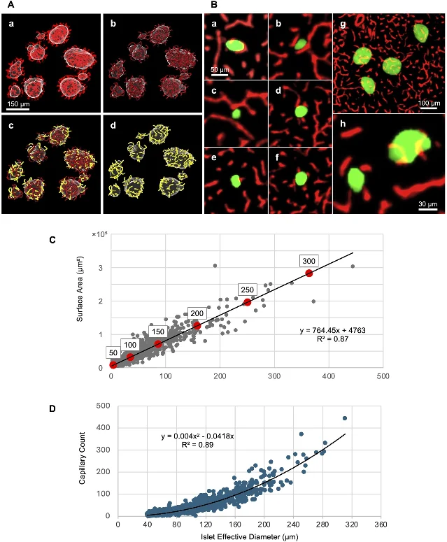
This is a Perspective, built on the authors' own three-dimensional imaging of pancreatic vasculature across mammalian species including humans, plus an internal comparison of 2D against 3D quantification on roughly 110 to 120 islets. The authors isolate individual capillaries that cross the digitally rendered boundary of an islet, count them, and plot that count against the islet's effective diameter — its diameter once the shape is normalised to a sphere. No new animals or experiments are reported; the argument assembles the group's earlier 3D measurements into a definition. 2
Key finding
The smallest endocrine cluster that receives its own internal vascularization measures about 40 µm in effective diameter. Above that size, capillaries cross into the cluster's interior and their number rises with the cluster's surface area; below it, endocrine cells sit as single cells or small groups next to capillaries without being internally supplied by them. The authors propose using that presence of internal vessels as the boundary between an islet (≥ 40 µm) and a non-islet single cell or small cluster, in place of the arbitrary size cutoffs different groups have used, which have ranged from 25 µm to 70 µm. 2
The paper then prices the measurement problem. On the same islets, 2D rendering underestimated islet effective diameter by 25.2% and intraislet capillary density by about 9% against the 3D values. Per individual islet the gap was wider: the discrepancy between the area-based and volume-based capillary density ran to 18.5% in either direction, because a single optical slice catches vessels as dots and short lines. Islet diameter in 2D errs one way only, since a section usually misses the largest central plane. 2
Why it matters
How you count endocrine clusters changes what you report. The islet equivalent used to dose a transplant is defined at 150 µm effective diameter, and the field's newest term for these structures — endocrine objects — was introduced precisely because labs disagree about where an islet begins. A definition tied to whether a cluster has its own blood vessels is measurable, and it changes the small end of the distribution: the single cells and small clusters that a size cutoff would sweep in as islets, and that carry no internal vasculature to keep them alive, drop out. That matters most for stem-cell-derived grafts, where differentiation typically yields a spread of cluster sizes and the fraction below 40 µm is exactly the fraction most likely to be lost after transplant. 2
Main limitation
The threshold rests on the authors' earlier study rather than on data new to this paper, and the authors say the value can vary a little around 40 µm between individual subjects and would benefit from being tested across more species. They restrict the definition to the mature adult pancreas deliberately; during fetal and early postnatal development the islet vasculature is still remodelling, and they note that this is one region where the rule does not apply as written. The extension to type 1 diabetes, where islets lose beta cells and shrink, is argued from preserved capillary networks in existing cohorts and awaits its own three-dimensional confirmation. A Perspective carries no search protocol and no new measurements, so the numbers above are the group's, reported rather than replicated. 2
3. Spheroid or organoid, same cells: matrix context and stimulation order decide the readout
Evidence class: Direct — vascular microphysiological systems
Authors, institution, date: Tali Zitman-Gal, Maria Sobolev, Anna Talash, Shelly Tartakover Matalon, Guy Topaz, Jacob Chen, David Pereg, Gloria Rashid, Naomi Nacasch, Arik Dahan, Mordechai Deutsch and Keren Cohen-Hagai; Department of Nephrology and Hypertension, Meir Medical Center, Kafr Saba, with collaborating clinical and academic centres in Israel; Journal of Vascular Research, advance article, 30 September 2026. 3
System or model
The device is a hydrogel microchamber array (HMC): a grid of small wells moulded in a hydrogel, into which endothelial cells are seeded and left to self-assemble. Cells cultured on their own form spheroids that have no surrounding matrix; cells seeded together with fibroblasts, in a chamber holding extracellular matrix, form organoids in which the matrix supplies a context the spheroid lacks. The authors tracked morphometric change over time — how far the structures expanded, how round they stayed — after exposure to high glucose, and after treatment with the fixed combination of a GLP-1 receptor agonist and insulin (Xultophy), varying which of the two stimuli came first. 3
Key finding
The platform produced spheroids and organoids reproducibly. Adding fibroblasts raised the organisation and structural complexity of the endothelium beyond what endothelial cells reached alone. Glucose made the organoids expand and lose circularity, a change the authors read as structural remodelling of the construct. The GLP-1 receptor agonist/insulin combination attenuated those glucose-driven changes and preserved organoid morphology, and its effect depended on the order in which glucose and the drug were applied, with only partial mitigation of the glucose effect. Organoids responded to the stimuli more strongly than spheroids from the same cells, which the authors attribute to the extracellular-matrix context. 3
Why it matters
This platform is a low-cost way to put endothelium under a metabolic insult and a drug, with a readout simple enough to run repeatedly. Its transferable result for graft work is the contrast inside it: two culture formats built from the same endothelial cells gave different answers, and the difference was the presence of matrix and fibroblasts. A group that screens a compound or a diabetic milieu against endothelium in a spheroid, and a group that runs the same screen in a matrix-embedded organoid, are running different experiments, and this paper shows the organoid is the more sensitive of the two. The sequence dependence adds a design variable that a single fixed protocol would hide. 3
Main limitation
Everything here is in vitro, and the cells are endothelial and fibroblast, with no pancreatic tissue and no endocrine cells, so what the organoids model is endothelial behaviour under a metabolic milieu of the heart-kidney-metabolic kind. The endpoints are morphometric — expansion and circularity — which capture the shape of the construct rather than its barrier function, its permeability or any flow through it; nothing in the accessible record applies shear or measures transport. The paper's own setting, cardio-kidney-metabolic disease, sets the glucose and drug concentrations, and a graft site would present a different milieu. The abstract is all that is available so far, so the effect sizes behind the direction statements are not in hand. 3
4. A curved-vessel chip built on an entry-level bioprinter
Evidence class: Direct — vessel-on-chip methods
Authors, institution, date: Robert S. Fischer, Paniz Rezvan Sangsari, Carey E. Dougan, Madison Daminato, Thomas Jones, Clare M. Waterman and Nicole Y. Morgan; Laboratory of Cell and Tissue Morphodynamics at the National Heart, Lung, and Blood Institute, and the Biomedical Engineering and Physical Science Shared Resource at the National Institute of Biomedical Imaging and Bioengineering, US National Institutes of Health; Journal of Visualized Experiments, methods article, 29 September 2026. 4
System or model
The protocol prints a sacrificial ink into a soft extracellular-matrix gel with an entry-level bioprinter, then dissolves the ink to leave an open, curved channel that human endothelial cells line. The channel is built to carry physiological flow rates, to admit permeability measurement and high-resolution light microscopy, and to hold a matrix of physiological stiffness. The target geometry is the vessel that bends: tortuous segments and aneurysmal bulges, the shapes where flow separates and the endothelium reacts to it. 4
Key finding
The method yields perfusable channels with curved and coiled geometry from a printer that costs far less than the specialised fabrication lines most curved-vessel chips have needed, and it keeps the build compatible with live endothelial imaging at high resolution. The paper presents the platform and its assembly; its abstract reports no measured values for shear, permeability or endothelial coverage. 4
Why it matters
Curvature is where a straight microfluidic channel stops predicting what endothelium does. Flow through a bend separates, the shear field turns non-uniform, and endothelial cells respond to that. A protocol that reproduces those geometries cheaply, with a compliant matrix instead of a rigid one, makes the curved case reachable for labs that could not previously build it. For a perfused graft device, the same technique offers a way to give a channel the bends and the soft wall that native vessels have, and the same sacrificial-ink step transfers to any hydrogel the lab already uses. 4
Main limitation
This is a methods paper in a video journal, so it reports how to build the platform; the abstract gives no numbers for the flow, the barrier or the cell layer. The model reproduces a vessel's geometry and its stiffness range; a printed channel still lacks the cellular wall of a real artery or vein, with its smooth-muscle layer, its fibroblasts and its perivascular matrix, so an experiment in it isolates the shape and the flow from the rest of the vessel's biology. 4
5. Blood-derived endothelial colony-forming cells make vessels that survive transplantation
Evidence class: Adjacent — prevascularization strategy for engineered tissue; no endocrine cells
Authors, institution, date: Yoshiya Asano, Yoshiko Tamai and Hiroshi Shimoda; Hirosaki University; Scientific Reports, 30 September 2026. 5
System or model
The authors built scaffold-free human vascularized artificial tissues (HVATs) by the cryopreserved cell accumulation method, which stacks cells coated in a nanofilm of extracellular matrix into a sheet without any scaffold. The cells were human adipose-derived mesenchymal stromal cells (hASCs) paired with endothelial colony-forming cells (ECFCs) drawn from two different sources: cord blood and peripheral blood. The constructs were followed in culture, then transplanted under the skin of nude mice and examined out to four weeks. 5
Key finding
Both ECFC sources formed vascular networks within four days of seeding and matched HUVEC-based constructs on two-dimensional vascular length density and area fraction. The two sources diverged in detail: cord-blood ECFC vessels showed incomplete endothelial closure and less perivascular differentiation of the stromal cells in culture, while peripheral-blood ECFC vessels were more continuous. After subcutaneous transplantation, cord-blood ECFC vessels formed continuous endothelial lumens lined by pericyte- or smooth-muscle-like cells and kept maturing over the four weeks; tissues built with peripheral-blood ECFCs also engrafted and formed human-derived vascular structures that contained host blood cells and were wrapped in perivascular cells. Profiling of angiogenesis-related factors in the culture medium showed distinct secretion patterns between the two endothelial sources. 5
Why it matters
A graft that arrives with its own vessels does not have to wait for the host to grow them in, and the endothelial cell that builds those vessels has to be obtainable. ECFCs from peripheral blood can be drawn and banked from the recipient, which makes them an autologous source for a prevascularized construct. This paper's contribution is the comparison: both blood sources produced human vessels that persisted in the host, carried host blood and acquired perivascular support, and the peripheral-blood source, less orderly in culture, produced the more continuous structures in vivo. 5
Main limitation
The tissue is adipose-stromal and it sits under mouse skin; there are no pancreatic or endocrine cells anywhere in the construct and no islet function to protect, so the result establishes an endothelial cell source and a prevascularization method rather than anything about a beta-cell graft. The endpoints describe vessel structure — lumens, perivascular cells, the presence of host blood — and the abstract carries no measure of what fraction of the construct the vessels actually perfuse. The host is immunodeficient, which removes the immune rejection that would be part of the clinical problem, and four weeks is the observed span. 5
Read the original article in Scientific Reports.
What this week changes
The microgel study and the islet-definition perspective meet at the same place from opposite ends. One supplies vessels to a graft by delivering a chemokine locally and reports the graft as better vascularized; the other says which structures in that graft count. Read together, they set the two ends of a measurement: what to deliver, and what to count once it has arrived. The gap between them is the number the microgel paper does not print — the vessel density or area that would let a reader compare its effect against the tungstate result of two weeks ago, which did print them.
The islet-lane picture for stem-cell-derived grafts stayed where it has been. No study in this window measured vascularization of stem-cell-derived beta cells or stem-cell-derived islets. The nearest items to that question are the 40 µm threshold, which bears on how many of a differentiated graft's clusters can hold their own vessels, and the ECFC tissues, which supply a vessel bed from the recipient's own blood. Both are steps toward the question rather than answers to it.
The two platforms settle variables on the fabrication side. The microchamber array reports that matrix and fibroblasts change what endothelium does under a metabolic insult, and that the sequence of exposure does too; the printed curved-vessel protocol reports that tortuous geometry and a compliant wall are now reachable from a cheap printer. Set beside the two islet items, they are the variables — the matrix around the endothelium, the shape of the channel, the source of the endothelial cell — that a perfused graft model has to fix before its results about a graft mean anything.
References
- 1
- 2
- 3
- 4
- 5
This story was produced automatically by a channel. One sentence is all it takes for Neodrop to keep producing for you.