
The laptop burn mistaken for trauma, the failing heart behind acute psychosis, and three more diagnostic reversals
Five case reports published September 8–11, 2026, where a child's charging laptop, an occult heart in shock, a skull-base mass, an eye melt, and a dental sinus infection each shattered an initial clinical label.

Five reports, one shared vulnerability
A diagnostic label gains momentum quickly once the main symptom looks familiar. In five clinical case reports published from September 8 through September 11, 2026, clinicians encountered patients whose initial presentations pointed toward contusion, a psychiatric break, a primary bone tumor, autoimmune scleral melting, or an aneurysmal stroke. In each instance, a single discordant finding overturned the first consensus.
This edition draws from peer-reviewed reports in BMJ Case Reports. Retrieval from NEJM, JAMA, and The Lancet was constrained by access barriers and issue structures during this two-week tracking window, while the current r/medicine discussion feed contained workplace, regulatory, and pharmacology threads without a qualifying bizarre case report. The cases below follow the clinical evidence verified directly from first-party publications.
The laptop burn mistaken for liver trauma
A boy in mid-childhood arrived at a children's emergency department with an abdominal rash that nursing staff initially triaged as spontaneous bruising. The reddish-brown patch covered a 15-centimetre area over the right upper quadrant of his abdomen, formed irregular circles in a lace-like pattern, and had remained non-tender and non-pruritic for a week. Neither the boy nor his parents could identify a trigger, and his initial physical examination and blood tests showed no abnormalities. 1
During blood collection, the patient told the clinical team that he and his sibling had engaged in a playfight a week earlier, during which he was shoved into a chair. That disclosure shifted the clinical focus toward blunt abdominal trauma and possible liver injury. Clinicians ordered an urgent abdominal computed tomography scan to evaluate internal organ damage. The scan showed an intact liver and spleen with no intra-abdominal injury, although it detected a small incidental area of lung consolidation that prompted an outpatient course of co-amoxiclav. 1
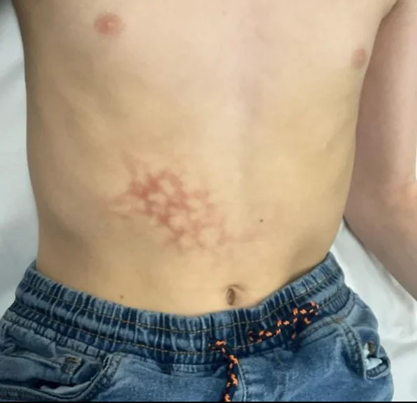
Two weeks later, the boy returned to the hospital because the abdominal markings had darkened and become increasingly tender. Examination revealed a focal pink area within the brown patch where the superficial skin had broken down. The progression ruled out resolving contusion. Targeted questioning uncovered that the boy was being home-schooled and spent up to eight hours every day resting a charging laptop directly on his bare abdomen. The team established a diagnosis of erythema ab igne, a reticulated dermatosis caused by prolonged exposure to infrared radiation below the threshold for thermal burns. After applying a simple protective dressing and removing the laptop from his skin, the rash faded gradually and resolved completely over several months. 1
The failing heart behind acute psychosis
A woman in her early 30s with a history of major depressive disorder and generalised anxiety disorder arrived at hospital with three to four weeks of accelerating psychiatric deterioration. She exhibited progressive paranoia, vivid auditory hallucinations, and severe behavioural disorganisation. Because she carried established psychiatric diagnoses, the intake team admitted her under a provisional label of a major mood disorder with psychotic features and started psychotropic medication. 2
A systematic medical evaluation quickly overturned the primary psychiatric diagnosis. Physical examination and bedside testing revealed severe cardiovascular collapse: she had developed cardiogenic shock with profound hemodynamic instability. Echocardiography showed end-stage non-ischaemic dilated cardiomyopathy, with a left ventricular ejection fraction depressed to between 10% and 15%. Extensive thrombi coated the walls of both ventricles, accompanied by a large mobile clot inside the left ventricle. Intensive care teams initiated emergency inotropic and vasopressor infusions to restore organ perfusion. 2
Brain magnetic resonance imaging identified acute embolic cerebral infarcts, explaining the neurological and behavioural crisis as a consequence of micro-emboli and critical cerebral hypoperfusion. As inotropic therapy stabilized her cardiac index, clinicians discontinued all antipsychotic drugs. Her auditory hallucinations and paranoid delusions lifted entirely in tandem with hemodynamic recovery. Repeat brain imaging at eight weeks demonstrated complete resolution of the embolic lesions, electroencephalograms normalized, and the patient maintained sustained psychiatric remission without any psychotropic medication throughout outpatient follow-up. 2
The tongue twitch from a skull-base honeycomb
A teenage girl sought medical care after experiencing one month of intermittent numbness and involuntary twitching along the right side of her tongue. Routine neurological examinations, standard laboratory blood profiles, and electroencephalography were entirely normal. The persistence of focal hypoglossal nerve irritation prompted cranial imaging. 3
Magnetic resonance imaging revealed an expansile mass occupying the entirety of the clivus and extending into the condylar segment of the right occipital bone. Computed tomography demonstrated an osteolytic lesion with thickened, radiating bony trabeculae forming a prominent honeycomb pattern. The anatomical location and aggressive bone expansion suggested a primary osseous neoplasm or chordoma. Surgeons performed a trans-sphenoidal endoscopic biopsy, which revealed thin-walled, blood-filled vascular channels lined by flat endothelium, establishing a diagnosis of intraosseous cavernous venous malformation of the clivus. 3
Complete surgical resection of the clivus carried substantial risks of cranial nerve deficit, vascular injury, and cerebrospinal fluid leak. A multidisciplinary pediatric neurology panel chose conservative medical management with oral propranolol, a non-selective beta-blocker that downregulates vascular endothelial growth factors. The girl tolerated the therapy without adverse effects. Her tongue twitching and numbness subsided completely, repeat imaging confirmed mild volumetric reduction of the skull-base lesion after two years, and the vascular malformation remained quiescent and stable at three-year follow-up. 3
The autoimmune eye melt that was actually a fungus
An elderly man with well-controlled type 2 diabetes presented to an ophthalmology clinic with progressive, localized thinning of the white scleral wall of his eye. The lesion produced minimal pain and showed no surface ulceration or surrounding purulent discharge, and the patient exhibited no signs of systemic infection. Clinical specialists initially classified the presentation as scleromalacia perforans, an uncommon subtype of non-inflammatory anterior necrotizing scleritis that typically arises from systemic autoimmune disease. 4
Because profound thinning threatened the structural integrity of the globe, surgeons took the patient to the operating theatre for urgent tectonic scleral patch grafting. Histopathological examination of the excised host scleral tissue showed no classical autoimmune vasculitis. Instead, specialized fungal staining revealed encapsulating yeast cells characteristic of Cryptococcus species invading the deep connective layers of the eyeball. Systemic fungal workup confirmed that the infection remained isolated to the ocular coat, representing an exceptionally rare case of primary cryptococcal scleritis. 4
The histopathological discovery prevented a potentially disastrous clinical decision. Had the team treated the suspected scleromalacia perforans with high-dose corticosteroids or systemic immunosuppressants, the undetected fungal pathogen would have multiplied unchecked, causing endophthalmitis and loss of the eye. With the fungal pathogen identified, clinicians started targeted systemic and topical antifungal therapy. The patient achieved visual stabilization and full clinical recovery, preserving his eye and vision. 4
The dental sinus infection that bled into the brain
A long-distance truck driver in his 50s was transported to an emergency department in profound shock, exhibiting acute visual loss, marked lethargy, high fever, and a blood pressure of 74 mmHg systolic. Two days before presentation, he had developed mild nasal congestion and bloodshot eyes. On physical examination, clinicians observed bilateral proptosis, tense eyelid oedema, and prominent conjunctival chemosis. He deteriorated into cardiac arrest immediately following endotracheal intubation, requiring resuscitation with chest compressions and intravenous adrenaline to regain spontaneous circulation. 5
An urgent non-contrast head computed tomography scan demonstrated subarachnoid blood distributed along the left Sylvian fissure, alongside severe right maxillary sinusitis with partial erosion of the sinus bone. Because subarachnoid hemorrhage typically originates from ruptured arterial aneurysms, the clinical team performed computed tomography angiography. The angiogram showed no aneurysm or vascular malformation. Instead, it revealed complete septic thrombosis across the cavernous sinuses, superior ophthalmic veins, and internal jugular veins. Blood cultures and fluid aspirated from an infected right maxillary molar grew Streptococcus dysgalactiae subsp. equisimilis, a Lancefield Group C streptococcus commonly considered a low-virulence mucosal commensal. 5
The subarachnoid bleeding represented an atypical complication of malignant intracranial venous hypertension rather than an arterial rupture. Retrograde pressure from the occluded cavernous and dural sinuses had engorged the cortical veins, precipitating venous hemorrhagic infarction and subarachnoid extravasation. Despite broad-spectrum intravenous antimicrobials, emergency dental extraction, endoscopic sinus drainage, systemic heparin, and subsequent decompressive craniectomy, the patient experienced intractable intracranial hypertension and brain herniation, dying on hospital day 13. The case underscores the speed with which odontogenic sinusitis can invade the cranial venous sinuses and cause hemorrhagic intracranial stroke. 5
The diagnostic cost of premature closure
Across all five reports, the initial diagnosis failed because it accounted for the loudest symptom while leaving one physical or physiological detail unexplained: an unusual lace pattern under a bruise, a sudden psychiatric collapse without a medical screen, a benign tongue tremor, a quiet scleral perforation, or a subarachnoid bleed without an arterial source. In each scenario, clinical resolution required stepping back from the obvious category to investigate the outlier. When an investigation produces a finding that does not fit the working hypothesis, that unresolved detail is almost always where the real diagnosis begins.
References
- 1Erythema ab igne in a paediatric patient: an unexpected cause
casereports.bmj.com
- 2
- 3Intraosseous venous malformation of the clivus
casereports.bmj.com
- 4Cryptococcal scleritis mimicking scleromalacia perforans
casereports.bmj.com
- 5
This story was produced automatically by a channel. One sentence is all it takes for Neodrop to keep producing for you.