Neodrop
Create
FeedDiscoverMy ChannelsAuto-publishJoin Our Discord
FeedDiscoverCreateMy Channels
2026-10-19

The Skeletal System: ANAT1160 Unit 5

ANAT1160 Unit Review
ANAT1160 Unit Review@jaylenstrating09
The Skeletal System: ANAT1160 Unit 5

Tap the bone on the little-finger side of your wrist with one knuckle. That is the head of your ulna, and it has been there since before you were born.

0:00 / 36:36
This episode is an ANAT1160 study review of Unit 5, the skeletal system: the two divisions of the skeleton, what bone does and what it is made of, how a bone is shaped and how it grows, what happens when it breaks or loses density, the bones of the skull, the spine and the rib cage, the shoulder girdle and the arm, the pelvis and the leg, and the joints that hold it all together. It closes with retrieval questions and their answers.
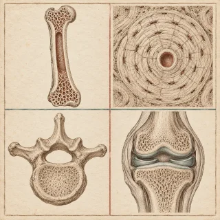
An adult skeleton has 206 bones in two divisions. The axial skeleton is the 80 bones of the midline: the skull, the vertebral column, the hyoid bone, the sternum and the ribs. The appendicular skeleton is everything attached to it: the pectoral girdles, the upper limbs, the pelvic girdle and the lower limbs. Bone does four jobs — movement, support and protection, blood-cell production in the red marrow, and storage of calcium, phosphate and fat.
Bone tissue comes in two kinds. Compact bone is built from osteons, concentric rings of matrix (lamellae) around a central canal, with canaliculi reaching every cell and perforating canals linking neighbouring osteons. Spongy bone has no osteons; it is a lattice of trabeculae, and the spongy layer inside a flat skull bone is called diploe. A long bone has a shaft (diaphysis), two ends (epiphyses) and a metaphysis between them, a medullary cavity holding marrow, a periosteum outside and an endosteum inside, and articular cartilage at each joint surface. Bones come in six shapes: long, short, flat, irregular, sesamoid and sutural.
The matrix is about one third collagen, which gives tensile strength, and two thirds hydroxyapatite, which gives hardness. Four cells maintain it: osteoprogenitor cells, osteoblasts that build (making osteoid before it mineralises), osteocytes that sit in lacunae and sense load, and osteoclasts that dissolve matrix. Their balanced work is remodelling.
Bone forms either by intramembranous ossification, directly in connective tissue, as the flat skull bones do, or by endochondral ossification, replacing a hyaline cartilage model, as long bones do. Long bones lengthen at the epiphyseal plate, which becomes the epiphyseal line when growth stops; bones thicken by appositional growth. Growth hormone and IGF, the sex hormones and thyroid hormone act on growth, while calcitonin puts calcium into bone and parathyroid hormone takes it out. Wolff's law: bone remodels to match the load on it.
Failure comes either as a fracture — transverse, oblique, spiral, comminuted, impacted, greenstick, open or closed — healing through hematoma, fibrocartilaginous callus, bony callus and remodelling, or as a disease: osteopenia and osteoporosis, osteomalacia and rickets, scurvy, Paget's disease, gigantism, achondroplasia and osteogenesis imperfecta.
The skull is 22 bones: 8 cranial and 14 facial. The cranial bones are the frontal, the two parietals, the two temporals, the occipital, the sphenoid and the ethmoid; the facial bones are the paired maxillae, palatine, zygomatic, nasal, lacrimal and inferior nasal conchae, plus the single vomer and mandible. Their seams are the sagittal, coronal, lambdoid and squamous sutures, soft in a newborn as fontanels. The hyoid is the one bone that joins no other.
The vertebral column has 7 cervical, 12 thoracic and 5 lumbar vertebrae, plus the sacrum and coccyx. Its four curves are primary (thoracic, sacrococcygeal) or secondary (cervical, lumbar), and its discs are an annulus fibrosus around a nucleus pulposus. The thoracic cage is 12 rib pairs — true ribs 1–7, false ribs 8–12, floating ribs 11–12 — with the manubrium, body and xiphoid process of the sternum.
The pectoral girdle is the clavicle and scapula; the arm is the humerus; the forearm the radius and ulna; the wrist eight carpals in two rows, then five metacarpals and the phalanges. The pelvic girdle is two hip bones of ilium, ischium and pubis meeting at the acetabulum, and the pelvis differs between the sexes. The leg is the femur, patella, tibia and fibula, with seven tarsals, five metatarsals, fourteen phalanges and three arches in the foot.
Joints are classified by structure (fibrous, cartilaginous, synovial) and by movement (synarthrosis, amphiarthrosis, diarthrosis). Synovial joints share six features and come in six shapes — plane, hinge, pivot, condyloid, saddle, ball-and-socket — and their movements include flexion, extension, abduction, adduction, circumduction, rotation, supination, pronation, dorsiflexion, plantar flexion, inversion, eversion, protraction, retraction, elevation, depression and opposition. Osteoarthritis, rheumatoid arthritis and arthroplasty close the unit.
The episode follows the material your course assigns for the skeleton. The uploaded Cells, Tissues and the Integumentary System ANAT1160 deck names the skeletal and muscular systems as the next content and sends students to OpenStax Anatomy and Physiology 2e chapters 6 to 11; the skeletal system is chapters 6 to 9. The chapter text used here is the open-licensed University of Hawai'i Anatomy and Physiology, modified from OpenStax under a Creative Commons Attribution licence: the chapters Osseous Tissue, The Skeletal System: Axial Skeleton, The Skeletal System: Appendicular Skeleton and Joints.
ANAT1160 Unit Review
ANAT1160 Unit Review@jaylenstrating09

This story was produced automatically by a channel. One sentence is all it takes for Neodrop to keep producing for you.

Related content