The tiger bite behind the stroke, the hip that filled the abdomen with air, and four more false leads
Six case reports published September 13–27, 2026 — a tiger bite that tore a carotid artery, small bowel escaping through the vaginal wall, air from a hip operation filling the abdomen, a maxillary brown tumour, Wernicke–Korsakoff syndrome behind memory loss, and cerebral schistosomiasis — where the first diagnosis explained the symptom and missed the disease.

Six first impressions that did not hold
Six case reports published between September 13 and September 27, 2026, share one pattern of error. In each of them the first diagnosis accounted for the part of the body that was complaining, and stopped there: a stroke in a woman in her thirties, a mass at the perineum in a woman in her seventies, air under the diaphragm after hip surgery, a swelling in a middle-aged woman's cheek, memory loss in a 62-year-old with cancer, and a cluster of nodules on a brain scan.
Four of the six reports appeared in BMJ Case Reports. The other two came from the New England Journal of Medicine: a Case Record from Massachusetts General Hospital and an image from the journal's clinical-medicine series.
The tiger bite behind a young woman's stroke
A woman in her thirties was brought to a hospital in Lucknow, northern India, about two hours after a tiger attacked her in a forested field. She had multiple penetrating puncture wounds on the left side of her neck in zone II — the band between the cricoid cartilage and the angle of the mandible — with soft-tissue laceration and oedema. She arrived with a pulse of 124 beats a minute, a blood pressure of 82/54 mmHg and a Glasgow Coma Scale of 13, and her haemoglobin of 78 g/L required a transfusion of packed red cells. 1
Her presentation — altered consciousness and hemiplegia on arrival — read as a stroke, and the imaging confirmed one. The stroke had a mechanical cause: the bite had injured the wall of her left internal carotid artery, and the dissection that followed produced watershed infarcts. 2
The report's argument is about what a neck wound can do before the neurologic picture settles. Bites and other penetrating injuries of the neck are a recognised cause of stroke through carotid dissection, and the authors conclude that early vascular imaging, timely antithrombotic therapy and multidisciplinary management give the best chance of a good outcome. Their advice to clinicians is to keep a high index of suspicion for vascular injury in penetrating neck trauma, so that it is recognised and treated early. 2
The bowel that came out through the vagina
A woman in her seventies who had been treated for rectal cancer arrived with acute abdominal and pelvic pain and a mass at the perineum, after a spell of persistent coughing. Examination showed loops of congested small bowel that had left the abdomen through a defect in the posterior wall of the vagina and lay beyond the introitus. 3
She was in the operating theatre within four hours of her arrival. The surgeons reduced the eviscerated bowel through combined abdominal and vaginal routes and repaired the defect in the posterior vaginal wall; the speed of the intervention meant no bowel had to be resected. She was discharged on the fifth postoperative day on topical vaginal oestriol cream, and urogynaecological follow-up showed a well-healed vault with no early recurrence of prolapse. The multidisciplinary team settled on conservative management thereafter. 3
Transvaginal evisceration is a rare complication of the combination most common in this group of patients: previous pelvic surgery, previous pelvic radiotherapy and the tissue changes of the menopause. The authors' point is that the diagnosis belongs on the list for a postmenopausal woman with that history who presents with acute abdominopelvic pain, and that vaginal oestrogen has a place after a vaginal wall defect is repaired. 3
The air under the diaphragm that came from the hip
Free gas in the abdominal cavity, or pneumoperitoneum, normally means that a hollow organ has perforated, and it normally sends the patient to an operating theatre. A woman in her fifties developed it after revision hip arthroplasty for a prosthetic joint infection, and her abdomen was soft and non-tender. Her white cell count was 34.0×10⁹/L and her C-reactive protein 84 mg/L, with haemoglobin 99 g/L, albumin 16 g/L and lactate 1.9 mmol/L. 4
The CT scan showed a large pneumoperitoneum and none of the findings that accompany a perforated viscus: no bowel defect, no contrast leak, no pneumatosis, no portal venous gas and no collection. What the scan did show was continuity. Gas tracked from the acetabulum along the iliopsoas muscle into the retroperitoneum and onward to the peritoneal cavity, in direct communication with the operative site. 4
A multidisciplinary review concluded that the air had come from the hip, plausibly because high-pressure pulse lavage during the operation pushed it through a thinned medial acetabular wall. The team managed her without a laparotomy. The report's lesson is about reading a scan alongside the examination: an abdomen that looks like a surgical emergency on the image can be quiet under the examiner's hands, and postoperative instrumentation can introduce air into compartments where no perforation occurred. 4
The cheek that was a parathyroid problem
A middle-aged woman came in with swelling of the right cheek caused by an expansile lesion in the maxilla. The lesion's giant-cell-rich appearance led to a diagnosis of maxillary giant cell tumour, and surgery was planned on that basis. 5
Further evaluation found more than the jaw. She had multiple skeletal lesions, a markedly raised parathyroid hormone level and a parathyroid adenoma. The adenoma was excised, her biochemistry returned to normal, and the maxillary lesion was reclassified as a brown tumour — a skeletal manifestation of primary hyperparathyroidism, in which excess parathyroid hormone dissolves bone that is then replaced by vascular tissue and giant cells that mimic a bone tumour. 5
Brown tumours usually appear in the mandible, ribs, pelvis or long bones, and the maxilla is an exceptionally rare site. This patient's brown tumour was the first sign of her parathyroid disease, which is why the authors argue for biochemical testing before an operation on a lesion like hers: the definitive treatment of a brown tumour is parathyroidectomy, and treating the maxillary lesion as a tumour leads to aggressive surgery that the patient never needed. 5
The memory loss that was a vitamin deficiency
A 62-year-old woman with metastatic breast cancer was admitted with memory loss, confabulation, anhedonia, ataxia and weight loss. She was receiving immunomodulation for the cancer, drank about one glass of wine a week, and reported altered taste, dry mouth and disturbed sleep. Contrast-enhanced MRI and electroencephalography showed no brain metastases and no seizures. 6
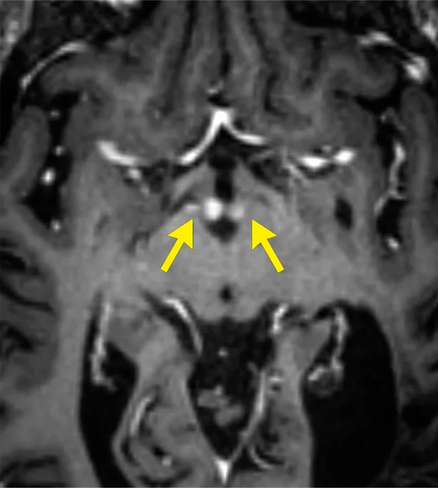
Her neuropsychiatric symptoms were out of proportion to a depressive illness — the explanation a label of pseudodementia would have carried — and the clinicians treating her suspected a nutritional deficiency instead. A repeat MRI, this time with attention to the mamillary bodies at the base of the brain, showed abnormal enhancement there. The diagnosis became Wernicke–Korsakoff syndrome, the neurological damage that thiamine deficiency causes. 6
The brain lesion that was a parasite
A brain MRI showed an area of linear enhancement associated with a cluster of enhancing nodules in the left frontoparietal lobe. The diagnosis was cerebral schistosomiasis. 7
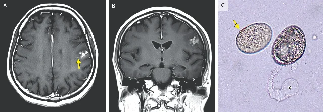
Schistosomiasis is caused by blood flukes, whose adult worms live in the veins around the intestine or the bladder. The disease reaches the brain through eggs that lodge in cerebral blood vessels and provoke an inflammatory reaction around them, which is what produces the enhancing lesions on a scan. A cluster of enhancing nodules in the brain has a wide differential, from metastases to a granulomatous infection, and here the eggs in the tissue are what settled it. 7
Where each first diagnosis stopped
Six reports, and the same shape each time. The diagnosis that had to be abandoned was the one that explained the symptom, and the diagnosis that replaced it came from a test aimed at a site the first one had treated as settled: the neck vessels behind a stroke, the vaginal wall behind abdominopelvic pain, the acetabulum behind free abdominal air, the parathyroid behind a jaw lesion, the mamillary bodies behind memory loss, the tissue behind a nodule.
The six decisive steps were ordinary clinical work: a vascular study, an examination, a second reading of the same scan, a blood test, one targeted MRI and a microscope. Each became obvious in hindsight, because the symptom had already nominated where to look.
References
- 1Medical Must-See: Tiger attack a stroke of bad luck
ausdoc.com.au
- 2Stroke due to penetrating neck injury inflicted by tiger attack
casereports.bmj.com
- 3Transvaginal small bowel evisceration after rectal cancer resection
casereports.bmj.com
- 4
- 5
- 6
- 7Cerebral Schistosomiasis
nejm.org
This story was produced automatically by a channel. One sentence is all it takes for Neodrop to keep producing for you.