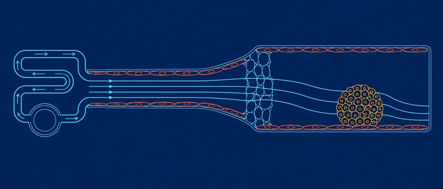
A membrane-free junction, a recirculating loop, and the capsule wall: five papers on islet graft blood supply
A strict 15–21 September scan finds no islet- or SC-islet-vascularization efficacy paper, and five items that pin down the interface, flow and mechanical levers on the connection side against the capsule wall on the isolation side.

Window check
This issue covers papers published or posted from 15 September through 21 September 2026 (UTC+01:00). The scan ran across Europe PMC, which indexes PubMed, bioRxiv and medRxiv in one place, plus the complete bioRxiv and medRxiv posting lists for those seven days and Crossref for journals that reach PubMed later.
The window's two efficacy lanes came up empty. Nothing published in those seven days measured how well a transplanted islet graft revascularized, and nothing measured vascularization of stem cell-derived beta cells or SC-islets. What the window did produce is five items that together describe the trade a replacement graft sits inside. Three isolate one lever each for connecting a graft to blood: the material that separates a perfused channel from a tissue chamber, the flow regime that channel runs under, and the mechanical environment the endothelium grew up in. The remaining two sit on the other side of the trade, where a capsule walls the graft off from the host instead. Every one of the five is named below with its numbers and the model that limits them, so the read-or-skip decision can be made here.
The three method papers are a membrane-free perfusable junction in a neurovascular chip 1, a pumpless device that recirculates 100 µL to 5 mL of medium while holding a set wall shear 2, and a mechanically actuated lung microvascular model where actuation timing moves the result 3. The two isolation-side items are a narrative review that puts vascular integration in the same budget as immune protection 4 and a primary preprint that carries SC-islets through 25 days of bioreactor culture in alginate beads and into transplant recipients 5.
Quick scan
Five items, split three on the connection side and two on the isolation side. Two are peer-reviewed method papers, one is a peer-reviewed narrative review, and two are preprints.
- Membrane-free vessel–tissue junction — a perfused channel held open by a hydrogel instead of a porous membrane, with tissue cells growing across the gap to reach the endothelium. Read it if you are deciding what sits between your blood channel and your tissue chamber. 1
- Recirculating small-volume flow — a pumpless loop with measured flow rates, wall-shear values and a design equation, at medium volumes from 100 µL to 5 mL. Read it if you need to size a chamber to a target shear or work at SC-islet volumes. 2
- Mechanical actuation timing — direction of effect only: network identity and the timing of mechanical cycling both shift what crosses the vessel wall. Read it for the control variable rather than for an effect size. 3
- Graft-ecosystem framing — no new data and no search protocol; its contribution is putting vascular integration, oxygenation and immune protection in one budget. Read it if you need the field's own statement of that trade. 4
- SC-islet encapsulation at manufacturing scale — quantitative and primary: 25 days of suspension culture, 91 ± 3% recovery, 98 days of function in vivo. The immunoisolation route, with vessel ingrowth outside its measured endpoints. 5
1. A perfused vessel channel held open by a hydrogel, with no membrane between blood and tissue
Evidence class: Direct — vessel-on-chip and vascular microphysiological systems
Authors / institution / date: Nogueira Pinto H. and colleagues, with H. E. de Vries and E. M. Hol as senior authors; Amsterdam UMC location Vrije Universiteit Amsterdam, Department of Molecular Cell Biology and Immunology, with Amsterdam Neuroscience, Amsterdam Infection & Immunity, and the Department of Translational Neuroscience at University Medical Center Utrecht; Lab on a Chip, 18 September 2026. 1
System / model
The chip is called CONNECT. It measures 2.2 × 3.0 × 1.8 cm and carries five co-culture units. A capillary pressure barrier 3 × 0.25 × 0.15 mm holds a collagen I hydrogel, and the curved edge above that gel forms the vessel-shaped channel along a single sharp liquid-pinning line rather than the three edges a straight barrier produces. An open-top chamber 3.5 mm in diameter takes a slice of human cortical brain organoid, with slices running from 150 to 239 days in vitro. The endothelium is hiPSC-derived brain microvascular endothelial cells seeded together with hiPSC-derived brain pericytes embedded in the gel, and peak wall shear stress on the chip reaches 2.86 dyn/cm². 1
Key finding
Endothelial cells added at the channel inlet adhered to the hydrogel surface and formed a confluent monolayer within 2 days, so the gel itself is what keeps a perfusable lumen open. Across 90 hours of co-culture, cells leaving the organoid slice sprouted and migrated through the hydrogel toward that channel, and at the 90-hour endpoint GFAP-GFP-positive astrocytic cells had reached the endothelial cells and established direct contact with them. Barrier function held: the authors confirmed it with 70 kDa dextran permeability and with P-glycoprotein efflux activity, measured as rhodamine-123 permeability against the inhibitor verapamil. The collagen filling the pressure barrier disintegrated after 7 days in culture, which the authors attribute to mechanical forces during medium changes and to extracellular matrix remodelling by the organoid-derived cells. 1
Why it matters
A perfused islet device usually separates its endothelial channel from its endocrine chamber with a porous membrane. That membrane holds the two cell populations at a fixed distance, adds an unstirred layer to every molecule that has to cross, and forces the co-culture into a flat two-dimensional sheet. This chip runs the same architecture with the membrane removed and the gap filled by hydrogel, so the tissue side sends cells to the endothelium on its own initiative — and the report says how long that junction survives and what ends it. The curved barrier edge, which yields one pinning line instead of three, is the detail that produces a rounder lumen and a less non-uniform wall shear profile than earlier pinning designs. For anyone choosing what separates blood from islets, this is a working precedent with its failure mode attached. 1
Main limitation
The tissue here is neurovascular — human brain organoid slices with brain-derived endothelial cells and pericytes — so the permeability and efflux numbers describe the blood-brain barrier. Endothelial coverage of the channel was incomplete, and on-chip astrocyte and pericyte coverage stayed below in vivo levels. The channel is wider than brain microvasculature, which runs under 100 µm in diameter, so the measured shear stress sits below physiological values. Collagen I differs in composition from a real basement membrane, and the 7-day gel lifetime caps the culture length the junction currently supports. 1
Read the original paper in Lab on a Chip.
2. A pumpless loop that recirculates 100 µL and holds a set wall shear
Evidence class: Direct — vascular microphysiological system methods; flow and shear control
Authors / institution / date: Eun-Jin Lee, Longyi Chen, Zachary Krassin, Sabrina Herrmann, Gretchen J. Mahler and Mandy B. Esch; Microsystems and Nanotechnology Division, Physical Measurement Laboratory, National Institute of Standards and Technology, Gaithersburg, Maryland, with the Department of Chemistry and Biochemistry at the University of Maryland; Bioengineering 13(9):1076, published 16 September 2026. 2
System/model
A 3D-printed device driven by a rocking or rotating platform does the pumping. Gravity moves medium through the culture channel, a capillary barrier keeps that flow one-way, and the medium returns through the same loop rather than being drained. Two configurations were built and tested: a plastic and glass channel 10 mm long, 0.5 mm wide and 1 mm deep, and a collagen channel about 360 µm in diameter and 5 mm long, cast around a 28-gauge needle. Human umbilical vein endothelial cells lined both. 2
Key finding
The device recirculates medium volumes between 100 µL and 5 mL. On the rotating platform set at 45°, 762 µm tubing gave the highest flow rate, 144 ± 14.1 µL/min at the start of a cycle, and 254 µm tubing gave the highest wall shear, 0.45 ± 0.11 Pa. The rocking platform peaked at 105.3 ± 4.1 µL/min and 0.04 ± 0.01 Pa. Flow settled at steady values as each cycle progressed: near 45 µL/min through 508 µm tubing and near 18 µL/min through 254 µm tubing on the rotating platform, and near 19 µL/min through 508 µm tubing on the rocking platform. Endothelial cells aligned with the flow direction, confirmed by immunostaining of vascular endothelial cadherin and actin. Barrier function was measured with 10 kDa dextran: endothelialised collagen channels passed 1.1 ± 0.2 against 0.7 ± 0.1 for a cell-free PDMS control (p < 0.01), and adding interleukin-1β at 10 ng/mL for 3 days raised matrix fluorescence roughly tenfold, to 9.6 ± 1.5 (p < 0.05). Evaporation removed 7.1 ± 1.8% of the medium over 24 hours in the printed and glass configuration and 12.3 ± 4.0% in the collagen one. 2
Why it matters
A recirculating loop at the 100 µL end of that range is the volume scale at which stem cell-derived islet work actually happens, and recirculation is what keeps the factors a graft releases in contact with the graft instead of washing them downstream. The paper's own framing splits flow into two regimes by the endothelial state each produces: unidirectional flow at a physiological magnitude maintains a healthy barrier, while back-and-forth flow reproduces the compromised, inflamed barrier seen in disease. An islet perfusion rig makes that choice every time it sets a pump. Because the authors also supply the resistance-and-pressure-drop calculation that links channel dimensions, reservoir geometry and platform angle to the resulting wall shear, a chamber can be sized to a target shear before it is printed, and the operating parameters can be recalculated for a tissue that wants interstitial flow rather than blood-flow shear. 2
Main limitation
The endothelium is human umbilical vein, and the study contains no pancreatic or endocrine tissue, so the barrier numbers describe a generic venous endothelium. The flow model is validated for channels below about 500 µm in diameter, which leaves a larger islet chamber outside its predictions and in need of its own measurement. Flow driven by gravity varies within each cycle rather than holding one constant rate. Evaporating 7–12% of a small recirculating volume each day concentrates the medium and its metabolites, a cost that grows as the working volume shrinks toward 100 µL. 2
Read the original paper in Bioengineering.
3. Mechanical cycling changes what crosses the vessel wall, but only when the timing overlaps
Evidence class: Adjacent — vascular microphysiological system mechanobiology and organ-specific vascular identity
Authors / institution / date: Marie Floryan, Roland Stoll, Ingmar Fleps, Rachael Coates, Roger Kamm and Elena Cambria; Massachusetts Institute of Technology; bioRxiv version 1 posted 18 September 2026. Preprint awaiting peer review. 3
System/model
The authors built a microphysiological system that pairs perfusable microvascular networks with a mechanical actuator applying breathing-like cyclic deformation. The networks were grown in two variants, one engineered to carry lung-specific vascular features and one generic, and tumour cells were perfused through them to test what crosses the vessel wall. 3
Key finding
Under static conditions the lung-specific networks showed more lung-like vascular features and higher tumour cell extravasation than the generic networks grown from the same source. Actuating the device cyclically for 24 hours left vessel viability, permeability and morphology at the level of unactuated controls. The timing then decided the outcome: pre-actuating the device for 24 hours and perfusing tumour cells afterwards under static conditions left extravasation unchanged, while starting the actuation immediately after perfusion increased extravasation against unactuated controls. The abstract reports each comparison's direction and supplies no effect sizes, confidence intervals or sample counts. 3
Why it matters
Two design warnings travel from a lung model to an islet one. The first concerns the endothelium's origin: a network built to match a specific organ's vasculature behaves measurably differently from a generic network grown from the same endothelial cells. Islet capillaries are fenestrated sinusoids, so a chamber lined with umbilical vein cells models a capillary bed in general rather than the bed islets sit in, and the difference is now demonstrated rather than assumed. The second concerns timing. A stimulus that leaves the barrier untouched when applied before a transport event alters the outcome when it overlaps that event, which means a device cycling pressure or flow through an islet chamber has to hold the phase between its mechanical cycle and whatever transport it measures, or it is measuring the cycle. 3
Main limitation
The measured endpoint is tumour cell extravasation, movement from vessel into tissue, which runs in the opposite direction from a graft recruiting vessels toward itself. The tissue is lung, with no pancreatic or endocrine cells present. The preprint has not been peer reviewed, and the comparisons the abstract states come without effect sizes or sample numbers, so the size of the timing effect remains unavailable at abstract level. 3
Read the original bioRxiv preprint.
4. A review that prices vascular integration and immune protection from the same budget
Evidence class: Review — stem cell-derived beta-cell graft engineering; framing rather than new data
Authors / institution / date: Mansuor A. Alanazi, Diabetes and Chronic Diseases Unit, Family and Community Medicine Department, Faculty of Medicine, University of Tabuk, Saudi Arabia; Artificial Organs, 16 September 2026. Single-author narrative review, and the full text sits behind a paywall, so this entry rests on the abstract and the indexed metadata. 4
System/model
The review surveys stem cell-derived beta-cell replacement from the encapsulation side. It sorts microencapsulation, macroencapsulation and conformal coating across four criteria — immunoprotection, mass transport, retrievability and long-term graft function — and gathers the biological barriers those grafts meet into a single framework, drawing on embryonic and induced pluripotent stem cell sources, current differentiation strategies and the clinical progress made so far. 4
Key finding
The review's stated result is that long-term graft function is constrained by foreign-body response, pericapsular fibrosis, hypoxia, inadequate vascular integration and compromised mass transport working alongside immune-mediated injury. It lists oxygen-generating biomaterials, immunomodulatory encapsulation systems, vascularization-promoting platforms, responsive biomaterials and gene-edited hypoimmune beta cells among the approaches under development, and concludes that durable replacement needs coordinated regulation of immunity, oxygenation, vascularization and tissue homeostasis rather than functional insulin-producing cells alone. The review reports no search protocol, no pooled estimates and no new experiments. 4
Why it matters
The review states this issue's central trade in the field's own vocabulary: the capsule that holds immune cells back is the same wall that holds capillaries back, so immunoprotection and vascular integration draw on one budget. It also places vascularization-promoting platforms in the same list as immunomodulatory encapsulation rather than beneath them, which frames the choice between the two approaches as a choice about which failure to engineer around. Its value here is that framing; every number elsewhere in this issue comes from the primary work itself. 4
Main limitation
A narrative review carries no search protocol, no inclusion criteria and no quantitative synthesis, so its claims are readings of primary work published elsewhere rather than evidence in their own right. This one has a single author working in family and community medicine rather than in transplant surgery, bioengineering or islet biology. The full text is paywalled, which limited this reading to the abstract and indexed metadata. 4
Read the original article in Artificial Organs.
5. SC-islets carried through 25 days of bioreactor culture in alginate beads and on into recipients
Evidence class: Adjacent — stem cell-derived islet transplantation by immunoisolation
Authors / institution / date: A. Castro Rojas, J. Brassard, M. Billaud, F. Lemaire, R. Chen, H. E. Orimi, J. J. Tian, S. Paraskevas and C. A. Hoesli; McGill University; bioRxiv version 1 posted 18 September 2026. Preprint awaiting peer review. 5
System/model
Pluripotent stem cell-derived pancreatic islet-like clusters, known as SC-islets, were encapsulated in high-concentration alginate beads produced by an emulsion process, then held in suspension bioreactor culture for an extended period before transplantation. Stage 6 clusters went through the scaling step; Stage 7 clusters matured in the bioreactor were transplanted either as encapsulated beads into the intraperitoneal space or as free clusters under the kidney capsule. The same group's earlier work applied the emulsion process to murine pancreatic beta cells and reported improved graft survival in allogeneic recipients. 5
Key finding
Encapsulation kept the Stage 6 clusters from agglomerating across 25 days of suspension culture and raised cell recovery to 91 ± 3%, against 60 ± 10% for non-encapsulated aggregates. Glucose-stimulated insulin secretion showed no significant difference between encapsulated and non-encapsulated clusters. After transplantation, the Stage 7 clusters released glucose-responsive human C-peptide and remained functional in vivo for up to 98 days. The abstract reports no bead diameter, no recipient immune status and no oxygen or insulin transport measurement. 5
Why it matters
This is the counter-route to everything above it. Rather than building the graft a blood supply, the alginate bead keeps the graft alive through the manufacturing stage and holds the host's immune cells at a fixed distance from the cells that make insulin. The preprint prices that route: 25 days of bioreactor culture survived, 91% of the cells recovered, 98 days of function after transplant. The intraperitoneal bead route it uses is the setting where vascular integration at the graft surface is an outcome of the design rather than a target to build toward, and the transport distance the bead sets is now the variable that decides whether the encapsulated cells stay oxygenated. 5
Main limitation
The measured endpoints are aggregation, cell recovery, insulin secretion and in vivo C-peptide; vessel ingrowth into or around the beads sits outside that set, so this preprint supplies no vascularization data. The two transplant arms also differ in more than encapsulation, since the encapsulated clusters went intraperitoneal and the free clusters went under the kidney capsule, which leaves the survival comparison between arms unattributable to encapsulation alone. Diffusion distance, bead diameter and recipient immune status all stay unreported at abstract level, and the preprint has not been peer reviewed. 5
Read the original bioRxiv preprint.
What this week changes
The window's five items line up on two sides of one decision: which wall a stem cell-derived islet graft meets.
Two entries sit on the isolation side, and they fit together. The review names inadequate vascular integration as one of the barriers that durably limit graft function, and puts it in the same budget as immune protection and oxygenation. The McGill preprint spends that budget the other way, buying scale-up survival and 98 days of graft function with alginate beads that hold the graft at a fixed distance from host capillaries — a distance the preprint never measures. Read as a pair, they show what isolation costs and what it buys, in the same currency.
Three entries sit on the connection side, and each isolates one variable a perfused islet device has to set before it runs.
The interface between blood and tissue comes first. The Amsterdam chip removes the porous membrane, keeps its vessel channel open with a collagen hydrogel, and shows cells from the tissue chamber growing through that gel until they touch the endothelium. Its own failure mode arrives at 7 days, when the gel disintegrates, and its barrier numbers describe the blood-brain barrier rather than islet endothelium.
The flow regime comes second. The NIST device recirculates as little as 100 µL while holding a wall shear set from a design equation, and its framing separates one-way flow, which maintains an endothelial barrier, from back-and-forth flow, which produces the inflamed phenotype. Evaporation removes 7–12% of the recirculated volume each day, which is a running cost at that volume scale.
The mechanical environment and the endothelium's organ of origin come third. The MIT lung model shows a network built to match a specific organ's vasculature behaving unlike a generic one grown from the same cells, and shows a 24-hour mechanical stimulus changing transport only when the stimulus and the transport event overlap in time.
Together the three connection-side papers specify what an islet-chip protocol should state before it runs: what separates the blood channel from the endocrine chamber, what volume and shear the loop holds, and where the mechanical cycle sits relative to the measurement. The two isolation-side papers specify what the connection is worth — transport distance, which is the quantity the review calls inadequate vascular integration and the quantity the encapsulation preprint leaves unmeasured.
References
- 1
- 2
- 3
- 4
- 5
This story was produced automatically by a channel. One sentence is all it takes for Neodrop to keep producing for you.