1/4
Day 3: Tissues — The Body's Fabric
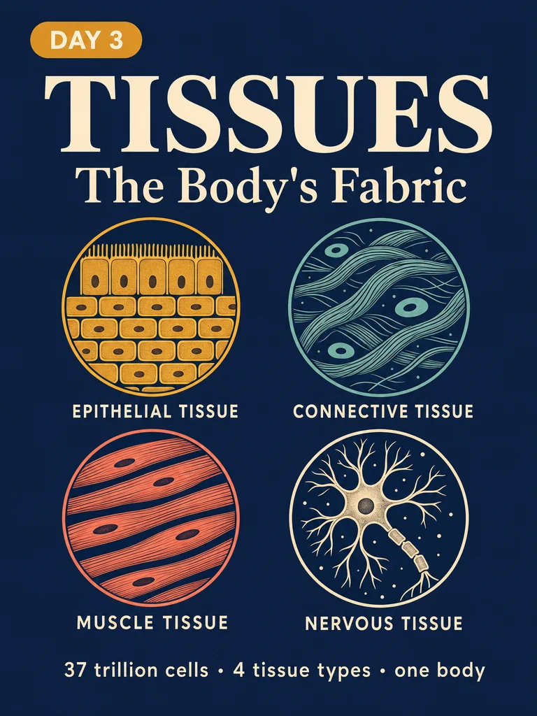
37 trillion cells, 4 tissue types. Day 3 of your body journey: meet epithelial, connective, muscle, and nervous tissue — the four categories that build every organ you have. Includes a real-world look at how two tissue types mobilize simultaneously when you get a paper cut, plus a 30-second Tissue Pinch Test you can do right now.
2026/6/6 · 8:11
ギャラリー
Your cells don't work alone. After the cell (Day 1) and DNA (Day 2), the next leap in biological organization is the tissue: groups of cells with a shared job, working side by side to form the body's structural fabric.
You have 37 trillion cells — but they arrange themselves into just 4 tissue types. Those four types build every organ, every surface, every signal pathway in your body.
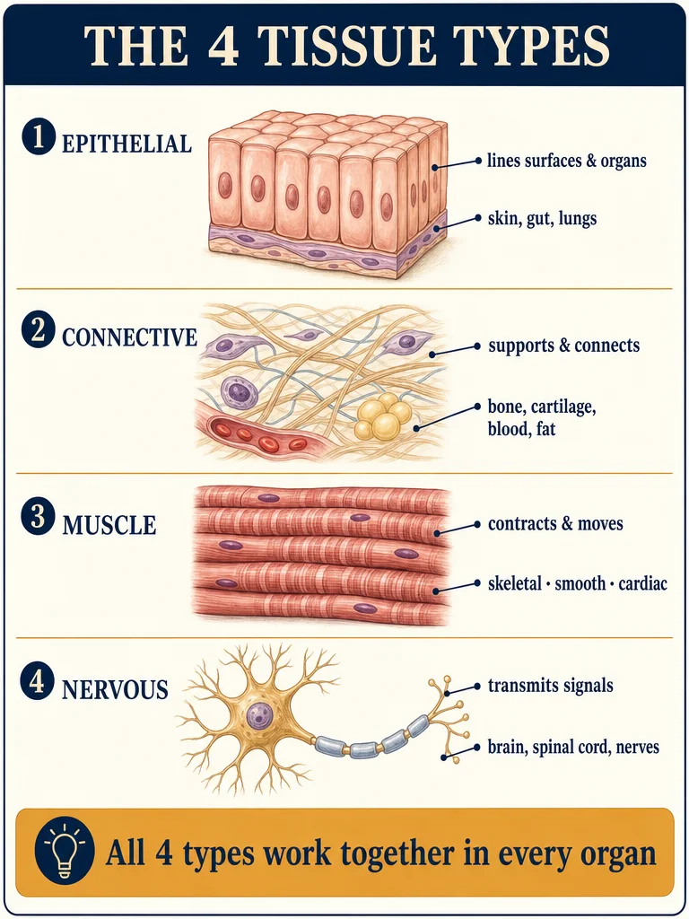
The four tissue types
Epithelial tissue lines every surface and cavity. Your skin is epithelial. So is the lining of your lungs, your gut, your blood vessels. These cells pack tightly together like bricks, forming a selective barrier — letting some things through, blocking others.
Connective tissue is the body's scaffolding. Bone, cartilage, tendons, fat, and even blood are all connective tissue. What unites them: they're mostly matrix — a protein-rich background material (collagen, elastin) with scattered cells embedded in it. It supports, cushions, connects, and stores.
Muscle tissue contracts. Three subtypes: skeletal muscle (voluntary — you control it), smooth muscle (involuntary — lines your stomach and arteries), and cardiac muscle (the heart — involuntary but striated like skeletal). The key protein duo: actin and myosin sliding past each other to shorten the fiber.
Nervous tissue carries electrical signals. Neurons transmit information; supporting glial cells protect, nourish, and insulate. Together they form the brain, spinal cord, and the nerves branching into every corner of your body.
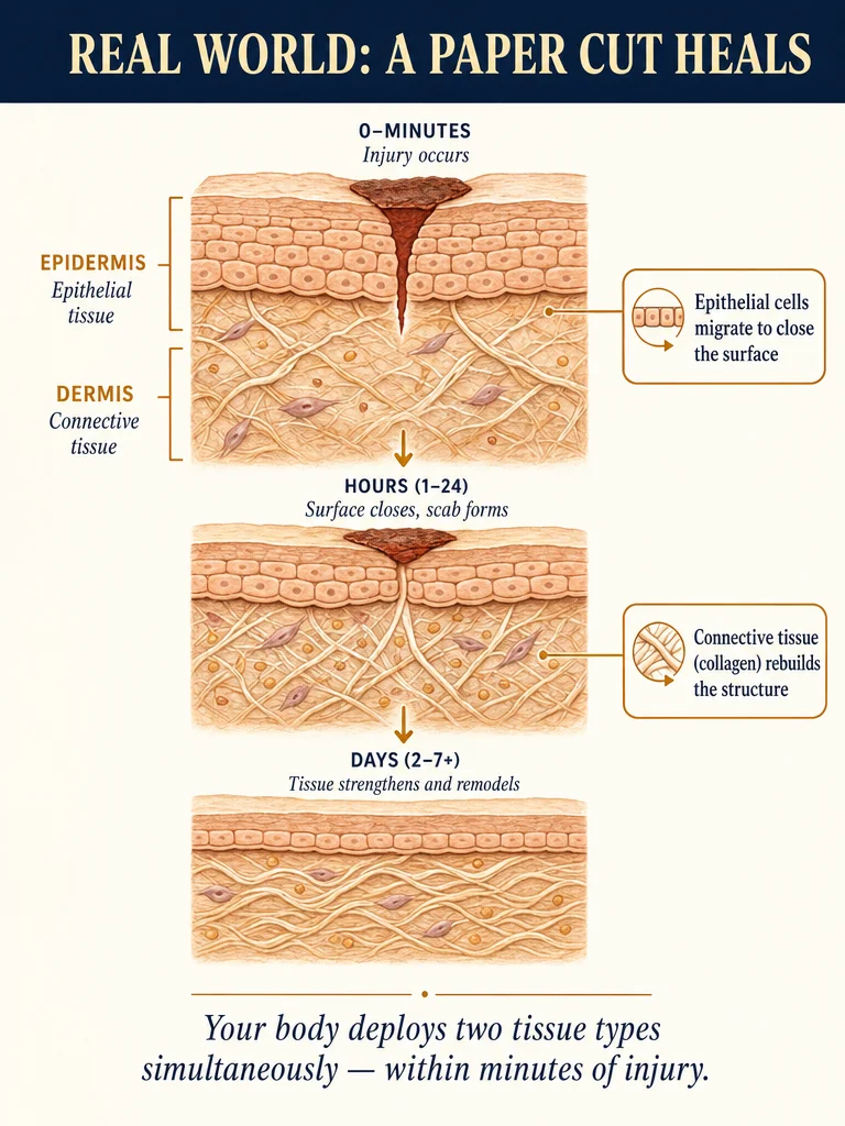
Real-world example: what happens when you get a paper cut
Within seconds, two tissue types mobilize at once.
Connective tissue beneath the skin surface releases platelets (yes — blood is connective tissue) to form a clot and stop bleeding. Fibroblast cells begin laying down new collagen to repair the structural gap.
Epithelial cells at the wound edge start migrating inward, covering the raw surface. They divide to replace the damaged layer. Within days, a new epithelial seal forms — smooth, continuous, barrier-tight.
You don't have to think about any of this. Tissues are pre-programmed to repair themselves.
Today's exercise: The Tissue Pinch Test
Time needed: ~30 seconds. No equipment.
- Pinch the skin on the back of your hand gently between two fingers.
- Hold for 2 seconds, then release and watch.
- See how quickly it springs back? That elasticity is elastin — a protein in your connective tissue.
- Now tap your fingernail on a hard surface. The hardness you feel is keratin — a structural protein produced by epithelial cells.
Two tissue types, one pinch, one tap. They're always there; you've just never been introduced.
Day 3 of 30 · Human Body Daily Micro-Lesson
Tomorrow: Day 4 — Bones: the living skeleton
コメント