The Fusiform Body Area: The Brain's Neighbor to the FFA
Three millimeters from the fusiform face area sits a completely separate region for perceiving human bodies as wholes — the fusiform body area (FBA). This article explains how Peelen & Downing found it, how Schwarzlose, Baker & Kanwisher named it with high-resolution fMRI, and why it differs fundamentally from the EBA.

The fusiform gyrus sits on the underside of the temporal lobe, folded away from view on the brain's ventral surface. For years, neuroscientists thought it had one job: recognizing faces. The discovery of the fusiform face area (FFA) in 1997 seemed to confirm this. Then came an awkward finding — when participants viewed headless human bodies, the fusiform gyrus lit up too, nearly as strongly as it did for faces. That activation turned out to belong to a previously unnoticed neighbor: the fusiform body area, or FBA.
The discovery: bodies in face territory
The FBA was first reported by Marius Peelen and Paul Downing in a 2005 paper in the Journal of Neurophysiology 1. Their experiment was simple but pointed. Participants in an fMRI scanner viewed images from four categories — faces, headless human bodies, tools, and scenes — while the researchers measured BOLD responses across the fusiform gyrus.
The result surprised the field. The mid-fusiform gyrus showed almost the same degree of selectivity for headless bodies as it did for faces, relative to the tool and scene baselines. A group analysis of 22 participants showed that the body-selective and face-selective fusiform activations were spatially overlapping at standard fMRI resolution (3.75 × 3.75 × 5.0 mm), but within-subjects analyses revealed that the peaks of the two activations sat in distinct but close locations. The face-selective peak — the FFA — fell somewhat more medially; the body-selective peak lay laterally adjacent to it.
A second experiment in the same study provided an important control. Peelen and Downing showed participants stick-figure depictions of human bodies alongside scrambled control images. The body-selective fusiform region responded more strongly to stick figures than to scrambled controls; the face-selective region did not. This mattered: it showed that the body response was not just a response to low-level visual features shared between bodies and faces, but generalized to abstract, schematic representations of the human form.
These results directly challenged the prevailing assumption that the fusiform gyrus was a face-processing region. It appeared instead to host multiple category-selective representations packed into adjacent columns of cortex.
Naming the region: Schwarzlose, Baker & Kanwisher (2005)
Shortly after Peelen and Downing's paper appeared, Rebecca Schwarzlose, Chris Baker, and Nancy Kanwisher at MIT formally named the body-selective region the fusiform body area (FBA) in a study published in Journal of Neuroscience 2.
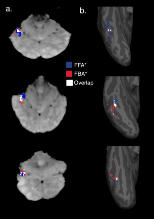
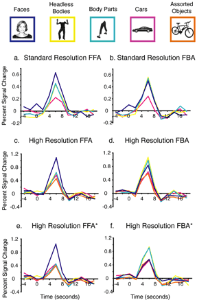
Their study addressed a methodological question: was the body response in the FFA a genuine body-selective signal, or did it arise from spatial blurring — signal from the FBA bleeding into the FFA at standard scanning resolution? They ran the same experiment at two resolutions: standard (3.125 × 3.125 × 4.0 mm) and high (1.4 × 1.4 × 2.0 mm).
At standard resolution, the results replicated Peelen and Downing — the FBA and FFA overlapped substantially, and both regions responded to both faces and bodies (though each more strongly to its preferred category). At high resolution, the picture changed. The dual selectivity weakened. When Schwarzlose et al. created "purified" ROIs by removing all voxels with overlapping selectivity — calling them FFA* and FBA* — the remaining regions showed exclusive selectivity: the FFA* responded selectively only to faces, not bodies; the FBA* responded selectively only to bodies, not faces.
The average distance between the centers of mass of the FFA* and FBA* was just 3.1 mm — a gap easily blurred by the interpolation and partial-voluming inevitable at standard fMRI voxel sizes. The apparent dual selectivity of the FFA for both faces and bodies, which had seemed to complicate the category-specificity argument, turned out to be a resolution artifact.
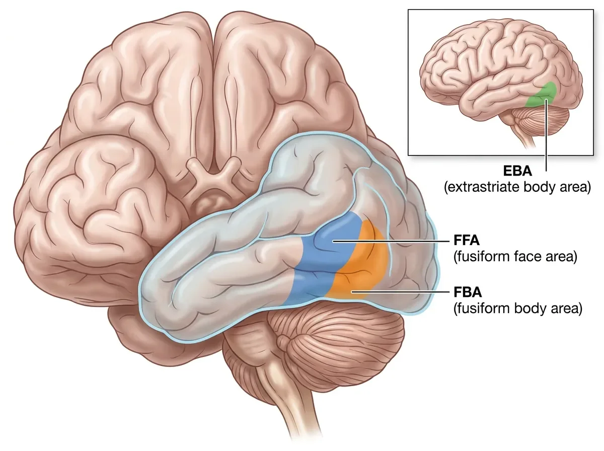
Location: neighbors on the fusiform gyrus
The FBA and FFA are both located in Brodmann area 37, in the mid-fusiform gyrus on the ventral (bottom) surface of the temporal lobe. The FBA sits on the lateral edge of the fusiform; the FFA sits slightly more medially, separated by just a few millimeters. Both are more reliably and more strongly activated in the right hemisphere, consistent with the right-lateralization seen in face processing more generally.
This positions the FBA in a very different part of the brain from the extrastriate body area (EBA) described by Downing, Jiang, Shuman and Kanwisher in 2001. The EBA sits on the lateral surface of the posterior temporal lobe (lateral occipitotemporal cortex), while the FBA is tucked into the ventral fusiform surface. They are centimeters apart and serve distinct roles, despite both responding to human bodies.
What the FBA does: holistic body representation
The core functional distinction between the EBA and the FBA became clearer in a 2007 fMRI study by John Taylor, Alison Wiggett, and Paul Downing 3. They systematically manipulated how much of the human figure was visible — from a single finger, to a hand, to an arm, to a torso — and measured the BOLD response in both EBA and FBA.
The EBA showed a gradual, linear increase in activation as more of the body came into view. More body = more EBA response, in rough proportion. The FBA showed something quite different: a step-like function. It was mostly unselective for individual fingers and hands. Activation jumped when a torso became visible and large body segments appeared. Once a substantial portion of the body was present, the FBA response reached its ceiling and stayed there.
This pattern mirrors the relationship between the occipital face area (OFA) and the fusiform face area (FFA) in face processing. The OFA shows incremental responses to individual facial features; the FFA cares about faces as unified wholes. Taylor et al. proposed the same architecture for bodies: EBA processes parts, FBA processes wholes.
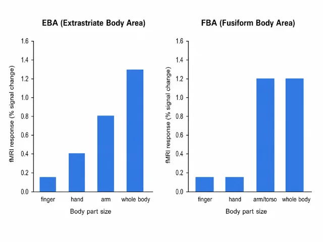
This part-vs-whole distinction has concrete implications. The FBA may be specifically involved in perceiving the overall configuration of the human body — the spatial arrangement of torso, limbs, and the body's overall posture and shape — rather than tracking individual components. The EBA, sitting earlier in the hierarchy, handles the parts.
Beyond the visual: abstract representations and known bodies
Several lines of research have extended the basic body-selectivity finding.
Abstract formats. The original Peelen & Downing paper already showed that the FBA responds to stick-figure bodies, not just photographic ones. This suggests the region is encoding something about human body structure — the spatial layout of limbs, the torso-limb configuration — rather than low-level visual properties. The same point is reinforced by the finding that the body-selective fusiform region responds to inverted bodies less than upright ones, paralleling the face inversion effect.
Known versus unknown bodies. The FBA can distinguish familiar bodies from unfamiliar ones. People can be recognized by their body alone (without a visible face), and some evidence suggests the FBA contributes to this person-identity signal 4. This connects the FBA to social cognition — the ability to recognize not just that a human body is present, but whose body it is.
Body image in anorexia nervosa. A 2013 study by Suchan and colleagues found reduced functional connectivity between the left FBA and EBA in patients with anorexia nervosa, compared to healthy controls. The degree of reduced connectivity correlated with the severity of body image distortion. The authors proposed that body perception depends on coordinated activity between EBA (which analyzes parts) and FBA (which integrates them into a whole), and that disruption of this communication contributes to the distorted body image characteristic of the disorder 5.
An open question: what exactly does "holistic" mean?
The EBA-parts / FBA-whole analogy is appealing, but "holistic body processing" remains somewhat underspecified compared to the corresponding literature on faces. For faces, holistic processing has been operationalized precisely: it means configural encoding that is disrupted by inversion, sensitive to spatial relationships between features, and impaired when faces are composited across different identities (the composite face effect). A parallel battery of tests for holistic body processing is less developed.
One important debate concerns whether the FBA is better described as a region that encodes body identity (who is that?) or body configuration (what is the overall posture and arrangement?). These are different computations, and the existing fMRI data do not cleanly distinguish them. Evidence from neurological patients with selective deficits in body recognition — analogous to prosopagnosia for faces — would be illuminating, but such cases are rare and their anatomical profiles remain poorly characterized.
The FBA's relationship to action recognition is also unsettled. Peelen and Downing's work has shown that body-selective cortex is involved in coding for action categories (recognizing whether someone is running or reaching), but how this subdivides between EBA and FBA remains a question.
Connecting the map
The FBA completes a picture that the previous three articles in this series have been building. The fusiform gyrus — once thought to be face territory — turns out to house two neighboring, category-selective regions: the FFA for faces and the FBA for bodies, separated by about 3 mm and distinguishable only at high scanning resolution. The EBA handles bodies too, but does so differently and from a completely different cortical location.
The organizational principle emerging from this work is a recurring one in cognitive neuroscience: the brain cares separately about the parts of an important category and the whole. We see it in vision for faces (OFA → FFA) and now in vision for bodies (EBA → FBA). Category-selective cortex is not a single detector; it is a processing hierarchy.
Landmark paper: Peelen, M.V. & Downing, P.E. (2005). Selectivity for the human body in the fusiform gyrus. Journal of Neurophysiology, 93(1), 603–608. PMID 15295012. 1
Naming paper: Schwarzlose, R.F., Baker, C.I. & Kanwisher, N. (2005). Separate face and body selectivity on the fusiform gyrus. Journal of Neuroscience, 25(47), 11055–11059. PMID 16306418. 2
Course connection: This concept maps to MIT 9.13 (The Human Brain) Lecture modules on face and body processing in high-level visual cortex, particularly discussions of category-selective regions in ventral temporal cortex and the debate between modular and distributed representations. The FBA sits directly in the territory explored by Kanwisher's lab over three decades of work on what the fusiform gyrus actually computes.
参考ソース
- 1Peelen & Downing, 2005 — Selectivity for the human body in the fusiform gyrus
- 2Schwarzlose, Baker & Kanwisher, 2005 — Separate face and body selectivity on the fusiform gyrus
- 3Taylor, Wiggett & Downing, 2007 — Functional MRI analysis of body and body part representations in the extrastriate and fusiform body areas
- 4Familiar body identity recognition in FBA
- 5Suchan et al. 2013 — Reduced connectivity between left FBA and EBA in anorexia nervosa
このコンテンツについて、さらに観点や背景を補足しましょう。