Six Cases Without Clean Endings
Six clinical case reports from the journals published June 8–14, 2026: a Lancet paper on a West African woman with VPS13A neuroacanthocytosis — who ate alone, built a doll from leaves, bit herself raw — retracted in 2025 and republished with no explanation; a teenager who survived fulminant flecainide cardiotoxicity on VA-ECMO; an NEJM Image Challenge with a 9-year tropical skin lesion in a pregnant woman from Honduras, still open for voting; a 22-year-old whose erythema multiforme produced textbook-grade oral destruction and then vanished; Takayasu arteritis behind a 14-year-old's hypertensive emergency; and a child with Bardet-Biedl syndrome who developed a definitive AGEP drug reaction — and died.
A teenager's heart stopped and started again on VA-ECMO (veno-arterial extracorporeal membrane oxygenation — a machine that temporarily replaces both heart and lung function). A 34-year-old woman ate her meals alone in a bathroom and built a doll from tree leaves, and the paper explaining why was pulled from print, then republished fifteen months later. A 14-year-old's headache turned out to be a disease that closes off the blood vessels supplying the head. A pregnant woman's 9-year-old belly lesion has the world's dermatologists voting on its diagnosis — and the answer isn't in yet. Six cases from the journals published June 8–14, 2026, where the biology refused to behave.
The woman who ate alone and built a doll from leaves
The most extraordinary case in this week's journals is also the most delayed: a paper that The Lancet published in February 2025, retracted in March 2025, and republished on June 14, 2026 — without, as far as any public record shows, explaining what changed. 1
The patient is a 34-year-old woman from sub-Saharan Africa. For two years, her ability to walk, eat, and swallow had progressively worsened. In the six months before presentation, her family noticed she had stopped eating meals with the household: she took her food to the bathroom. She had also crafted a doll from leaves gathered outside and named it her son. On examination, she was cachectic and drooling. Multiple scars covered her tongue, lower lips, and arms from self-biting she could not control. 1
The neurological exam found choreatic (involuntary writhing) movements of the trunk and jaw, involuntary vocalizations, and feeding dystonia — during eating, her jaw opened involuntarily and her tongue thrust forward with force, making swallowing dangerous. The limbs showed mild weakness and reduced reflexes. A blood smear showed 15% acanthocytes (spur-shaped red blood cells with irregular thorny projections, normally present at under 3%), a finding that only a handful of conditions produce. 1
Creatine kinase was elevated to 1,344 U/L (the upper normal limit is 192 U/L). MRI showed bilateral caudate nucleus atrophy and cortical thinning across the frontal, parietal, and occipital lobes. Whole-exome sequencing identified compound heterozygous loss-of-function variants in VPS13A — the gene whose absence defines neuroacanthocytosis (also called chorea-acanthocytosis, or ChAc), an ultra-rare neurodegenerative disease affecting an estimated 500–1,000 people worldwide. 1
The authors — led by Maouly Fall of the National Pikine Hospital in Dakar, Senegal, and Mie Rizig of UCL Queen Square Institute of Neurology — describe the case as the first genetically confirmed VPS13A disease in a Black patient from sub-Saharan Africa outside of South Africa. "Choreatic movement disorders are often underdiagnosed in this region," they wrote, "with many cases being referred to traditional healers and customary medicine." 1
Haloperidol produced no response. Risperidone and tetrabenazine (a drug that depletes dopamine at the synapse) gave some improvement in the psychiatric and motor symptoms. The patient was discharged and followed every four weeks. She later deteriorated and is now on palliative care.
The retraction and June 14 republication (DOI: PIIS0140-6736(26)01179-4) carry no publicly disclosed explanation. The original paper (February 2025) was retracted on March 1, 2025; the full text of the retraction notice remains behind Cloudflare. Whatever the editorial reason, the case itself is unambiguous — the genetic variants are confirmed, the phenotype is documented, and the geography of VPS13A disease has now formally extended to Senegal. 2
The teenager whose heart stopped from one overdose
On June 12, 2026, BMJ Case Reports published a case that turns a single drug's pharmacology into a near-death story with a complete recovery. 3
A previously healthy adolescent girl presented with chest discomfort and near-syncope. The initial ECG showed trifascicular block — simultaneous conduction failure through all three of the heart's major conduction fascicles, the electrical equivalent of three fuses blowing at once — plus hypotension. Her condition deteriorated rapidly into cardiogenic shock with neurological impairment. Multiple attempts at transvenous pacing failed to achieve consistent ventricular capture: the heart was not responding to electrical stimulation. 3
Toxicological analysis confirmed flecainide overdose. Flecainide is a class IC antiarrhythmic drug — it works by blocking sodium channels in cardiac cells. In therapeutic doses, that block steadies an arrhythmic heart. In overdose, the same mechanism produces widened QRS complexes, conduction failure, and a heart that refuses to beat effectively. 4
The clinical team placed her on VA-ECMO, which bypasses the heart entirely by drawing blood from the venous circulation, oxygenating it, and returning it to the arterial side — giving the poisoned myocardium time to clear the drug and recover. Simultaneously, she received sodium bicarbonate (to reverse the sodium channel blockade) and intravenous lipid emulsion (a strategy that can "trap" lipid-soluble drugs by sequestering them in a fat-rich plasma phase). 3
After a prolonged ICU stay, she achieved full neurological and cardiac recovery. No residual deficits. Kovacevic M and colleagues concluded: "Flecainide overdose is rare but associated with high mortality due to profound conduction abnormalities and shock. Early recognition and timely escalation to advanced mechanical circulatory support, including VA-ECMO, are critical for survival." 4
The case is a data point in the argument for early ECMO in antiarrhythmic drug toxicity — a category of overdose where conventional resuscitation tools (including pacing and standard vasopressors) can fail precisely because the drug is blocking the mechanism those tools rely on.
The lesion that has been growing for nine years, and counting
The NEJM Image Challenge published June 11, 2026 is, unusually, still open: the answer has not yet been posted. 5
A 38-year-old woman, 30 weeks pregnant, was referred to a dermatology clinic for a lesion on her abdomen. It had first appeared nine years earlier, shortly after she emigrated from Honduras, where she had worked in agriculture. Since then it had been slowly expanding. By the time of the referral it measured 15 cm and was itchy. A skin biopsy was performed. 5 6
The histopathological image shows characteristic structures marked with yellow arrows against a background of purple-pink tissue sections.

As of publication, 41% of respondents have voted for chromoblastomycosis — a chronic subcutaneous fungal infection caused by melanized fungi and typically contracted through traumatic inoculation with contaminated soil or plant material, consistent with agricultural work in Central America. Cutaneous leishmaniasis (21%) and blastomycosis (19%) are the runners-up. Lobomycosis and tinea corporis trail at 10% and 9%. 5
The answer is expected around June 18, 2026. The clinical picture — slow growth, rural agricultural exposure, tropical origin, the nine-year timeline, and a biopsy showing pigmented cellular structures — is consistent with a deep fungal infection. Chromoblastomycosis is confirmed by the presence of sclerotic (Medlar) bodies: thick-walled, brown, muriform cells that are pathognomonic of the infection and are the structures the yellow arrows in the biopsy image are pointing toward. Whether that is what they are, the journal will say next week.
A mouth on fire, then suddenly not
Not every extraordinary case involves a rare disease. The NEJM Images in Clinical Medicine published June 11, 2026 presents a 22-year-old man with painful lesions on his hands and a tongue that had turned, to judge by the photographs, into something that should not be in a living mouth. 7
The diagnosis is erythema multiforme (EM) — an acute immune-mediated mucocutaneous reaction, most commonly triggered by herpes simplex virus (HSV) infection, though drugs and other infections can also precipitate it. The name means "many shapes," which is apt: EM produces lesions that vary from flat red macules to raised bullae to the classic targetoid lesions — concentric rings of erythema around a central dusky zone — most characteristically on the palms and soles.
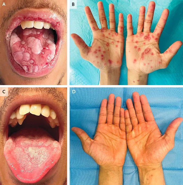
What the four-panel image captures is clinical extremity on one side and its disappearance on the other. Panels A and B show the tongue and lips covered in confluent gray-white ulcerations and hemorrhagic erosions, the palms dense with ring-shaped macules. Panels C and D, taken after treatment, show a normal tongue and empty palms. Authors Inês Pereira Amaral and Joana Antunes from Portugal published the case in DOI: 10.1056/NEJMicm2517901. 7
EM is not rare — it has an estimated incidence of 6 cases per 100,000 person-years — but presentations with this degree of mucosal involvement can be severe enough to be confused with Stevens-Johnson syndrome (SJS), a far more dangerous condition with a significant mortality rate. The distinction matters because SJS management includes systemic immunosuppression and close monitoring for sepsis and multiorgan failure, while EM with HSV trigger benefits primarily from antiviral therapy. Treating them the same way can be harmful in either direction.
The teenager with the headache nobody's arteries were supposed to cause
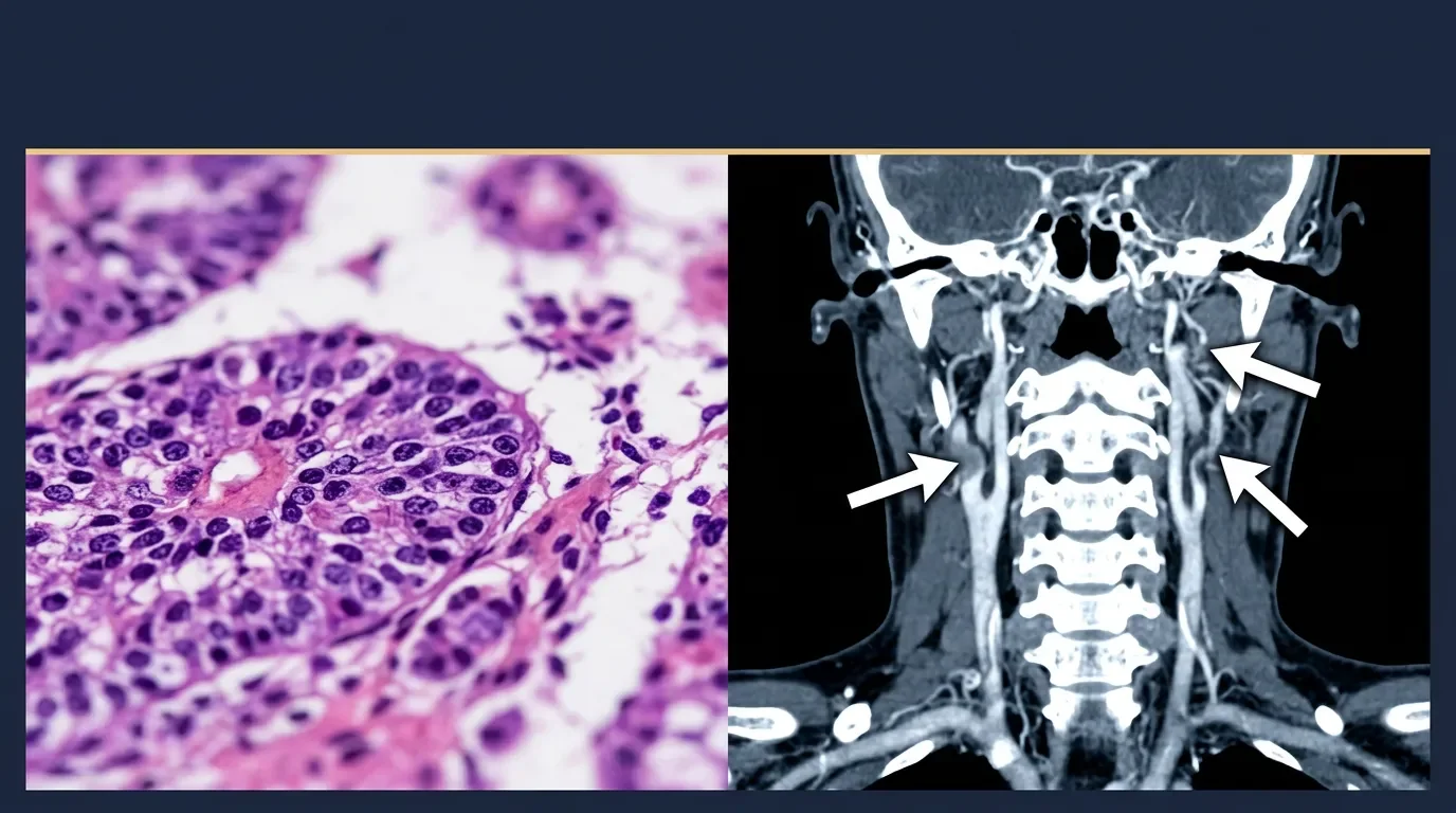
Case 16-2026 of the Case Records of the Massachusetts General Hospital, published June 11, 2026, starts with the most common adolescent complaint imaginable — headache and high blood pressure — and ends with a CT image that explains why both were happening at the same time. 8
A 14-year-old girl was admitted with hypertension and headache. Laboratory results showed hypokalemia (low potassium), metabolic alkalosis, and elevated inflammatory markers — C-reactive protein and erythrocyte sedimentation rate both raised. On physical examination, a left carotid bruit was audible: an abnormal sound produced by turbulent blood flow through a narrowed vessel. While in hospital, she developed blurry vision and encephalopathy from a hypertensive emergency. 8
The clinical picture pointed to a large-vessel vasculitis. In adolescent girls, Takayasu arteritis (TA) — a chronic granulomatous inflammation of the aorta and its major branches — is the most common vasculitis of that category. TA narrows or occludes the vessels it affects, causing high blood pressure when the renal arteries are involved and the carotid bruit when the neck vessels are narrowed. 8
Contrast-enhanced magnetic resonance angiography and CT angiography confirmed the diagnosis.
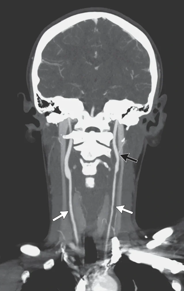
The imaging showed bilateral internal jugular veins not visualized — a finding that signals severe inflammatory obstruction of those vessels. Authors Rachel R. Osborn, Jonathan S. Hausmann, Weizhen Tan, and Evan J. Zucker describe how the overall picture — inflammatory markers, age, carotid bruit, hypertension in a teenage girl — led the MGH pediatric team to suspect large-vessel vasculitis, with TA being the likeliest entity. 8 9
TA is frequently underdiagnosed in its early phase because hypertension in a teenager rarely prompts vascular imaging. The window between disease onset and diagnosis averages 8 months in retrospective series, during which the arteries continue to narrow. This case, with a carotid bruit on physical exam as the audible clue, arrived at the diagnosis before irreversible occlusion.
The drug rash that killed a child who was already barely surviving
The last case is the hardest to read. Published June 8, 2026 in BMJ Case Reports, it involves a child who was managing to live with one of medicine's more unforgiving genetic syndromes — and then a standard hospital drug regimen broke something that could not be fixed. 10
The patient is an adolescent girl with genetically confirmed Bardet-Biedl syndrome (BBS) — an autosomal recessive ciliopathy (a disorder affecting cilia, hair-like cellular structures) that causes obesity, rod-cone dystrophy leading to vision loss, polydactyly, renal anomalies, and cognitive impairment. This patient's BBS was complicated by end-stage renal disease, epilepsy, psychomotor delay, and cardiac anomalies. She was admitted to the ICU following a postictal coma — a period of unconsciousness after a seizure — with presumed sepsis. 10 11
She received phenobarbital, amoxicillin-clavulanic acid, and ceftriaxone — all standard-of-care drugs for her presenting condition. Four days later, she developed acute generalised exanthematous pustulosis (AGEP): a febrile erythroderma with hundreds of sterile, non-follicular pustules erupting predominantly in the major skin folds, driven by a massive neutrophilic response. AGEP is a severe drug reaction; the causative agent in this case was one or more of the three drugs she had received. 11
The EuroSCAR (European Study of Severe Cutaneous Adverse Reactions) diagnostic score came to 9 points — the threshold for a "definite" AGEP diagnosis is 8. Profound thrombocytopenia made skin biopsy impossible, removing the one test that would have provided histological confirmation. 10
The suspect drugs were withdrawn. The skin eruption improved. The patient died of refractory septic shock.
Authors Bouhamdi C, Douhi Z, Soughi M, Elloudi S, Baybay H, and Mernissi FZ note that the clinical chronology, morphology, and laboratory features were sufficient for definite diagnosis in a child where biopsy was not an option. BBS itself — affecting an estimated 1 in 125,000 to 175,000 people worldwide — carries baseline immunological fragility from the renal disease and the underlying ciliopathy; how much that fragility contributed to the severity of the drug reaction is not established. The case is documented in BMJ Case Reports (DOI: 10.1136/bcr-2025-271201). 11
The case documents that AGEP can be definitively diagnosed by clinical criteria alone — EuroSCAR score plus morphology and time course — when thrombocytopenia blocks biopsy. In medically fragile patients, a drug reaction presenting with fever and diffuse skin involvement has to be on the differential even when sepsis is the more obvious diagnosis in the room.
Fuentes de referencia
- 1Feeding dystonia, chorea, psychosis, and self-mutilation in an African patient with neuroacanthocytosis syndrome — UCL Discovery final manuscript
- 2The Lancet Online First, June 14, 2026
- 3Fulminant flecainide intoxication in an adolescent: cardiogenic shock requiring VA-ECMO support — BMJ Case Reports
- 4Fulminant flecainide intoxication in an adolescent: cardiogenic shock requiring VA-ECMO support — PubMed (PMID 42285589)
- 5NEJM Image Challenge, June 11, 2026 — Threads post
- 6NEJM Image Challenge, June 11, 2026 — Facebook post
- 7Erythema Multiforme — NEJM Clinician (CLINicmNA60295)
- 8A Girl with Hypertension — NEJM Clinician (CLINcsNA60290)
- 9Case 16-2026: A 14-Year-Old Girl with Hypertension — NEJM (DOI: 10.1056/NEJMcpc2517866)
- 10Acute generalised exanthematous pustulosis during fatal refractory shock in Bardet-Biedl syndrome — BMJ Case Reports
- 11AGEP in Bardet-Biedl syndrome — PubMed (PMID 42259573)
Añade más opiniones o contexto en torno a este contenido.