The Occipital Face Area: How the Brain's Face Network Actually Works
The face-processing network is not a single area but a three-node system — OFA, FFA, and pSTS. This article explains what the Occipital Face Area is, where it sits in the hierarchy, and why the most striking finding in this literature is that removing it surgically leaves the downstream FFA almost entirely intact.

When we last visited the fusiform face area, we left with a clean story: a patch of cortex on the fusiform gyrus fires reliably to faces, and damage or disruption to it impairs face recognition. Tidy. But the FFA does not work alone. It sits at the middle of a three-node network that also includes a region most people have never heard of — the Occipital Face Area (OFA) — and a strip of cortex along the superior temporal sulcus. How these regions interact, whether they form a strict processing hierarchy or something messier and more interesting, is one of the more productively contested questions in face-perception neuroscience.
What the OFA is and where it lives
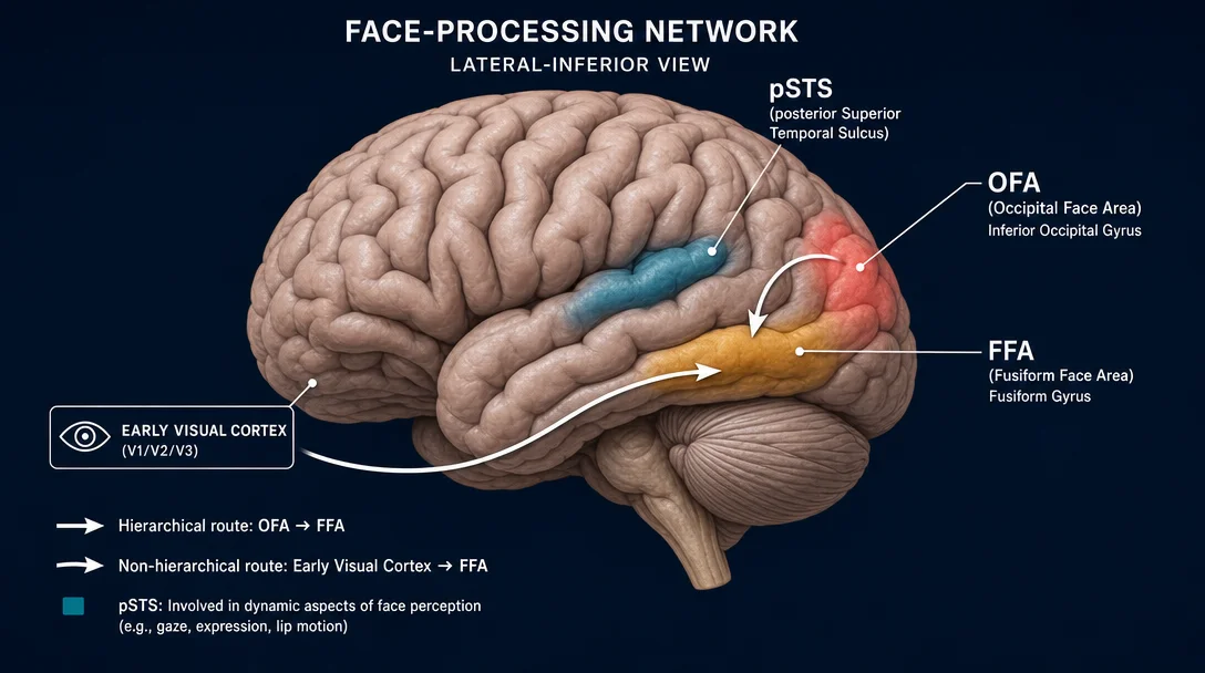
The OFA sits in the inferior occipital gyrus (IOG), on the lateral face of the occipital lobe, posterior to and below the FFA. In most people it sits at roughly MNI coordinates (±44, −76, −13) — further back in the visual cortex than the FFA, which anchors around the mid-fusiform gyrus. Like the FFA, it is bilateral but strongly right-lateralized.
The name was introduced by Gauthier et al. in 2000, in a Nature Neuroscience paper using fMRI to examine brain responses to newly learned objects. The key observation: the IOG, like the FFA, showed elevated activation not just for faces but for objects subjects had been trained to distinguish at a fine-grained level — suggesting this region is involved in expert-level visual differentiation, with faces being its most practiced domain 1. Whether the OFA is really "face-specific" or more broadly "expert-level discrimination" remains a live argument, but from that 2000 paper onward, "OFA" stuck as the shorthand for the face-selective patch in the IOG.
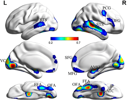
In standard face-localizer fMRI experiments — where subjects passively view faces alternating with objects, scenes, or scrambled images — the OFA activates consistently as one of three cortical regions forming what James Haxby and colleagues described in their influential 2000 Science paper as the core face-processing network: OFA, FFA, and the posterior superior temporal sulcus (pSTS) 2.
The Haxby model: a three-station hierarchy
Haxby's 2000 framework proposed a conceptually clean division of labor. The OFA handles the initial analysis of facial parts — the low-level geometry of eyes, nose, mouth, and their spatial layout. That information feeds forward to two downstream recipients:
- The FFA, which processes relatively invariant aspects of a face — the stable features that let you recognize a person regardless of viewpoint, expression, or lighting. This is the "who is this?" computation.
- The pSTS, which processes changeable aspects — expression, gaze direction, lip movement, head orientation. This is the "what is this face doing right now?" computation.
The hierarchy runs posterior to anterior and posterior to dorsal: OFA first, then a fork to FFA (ventral, fusiform cortex) and pSTS (more dorsal, temporal cortex).
The model was influential for good reasons. It unified a scattered fMRI literature under a single architecture, made testable predictions about what disrupting each node should do, and meshed with the broader logic of the ventral visual stream, where early areas extract local features and downstream areas integrate them into object representations. In this reading, the OFA is the face network's V1: the entry point that everything else depends on.
TMS evidence: the OFA and facial parts
One prediction follows directly from the hierarchy: disrupt the OFA and you should impair facial feature processing. That prediction has largely held. Transcranial magnetic stimulation (TMS) studies have repeatedly shown that applying a brief magnetic pulse over the OFA — timed to the presentation of a face — selectively disrupts the ability to discriminate individual facial features (eye shape, mouth configuration) without impairing holistic face processing or scene perception. TMS over the FFA produces the opposite pattern: impaired recognition of whole faces, less effect on isolated features 4.
This double dissociation — OFA for parts, FFA for wholes — was clean enough to be cited as strong causal evidence for the hierarchy well into the 2010s. It fit the computational story: parts come in at the back, integrated wholes emerge further forward.
The challenge: Patient PS and the FFA without OFA
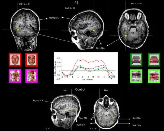
The trouble for strict hierarchy began with a single patient. Patient PS is a Belgian woman who sustained a closed-head injury in 1992, leaving her with extensive damage to the right inferior occipital cortex — the territory of the OFA — plus the left fusiform gyrus. She developed severe acquired prosopagnosia: she could not recognize her family members' faces out of context.
That part fits the hierarchical story. What does not fit is what happened when Bruno Rossion's group scanned her brain. Despite the lesioned right OFA, Patient PS showed normal face-selective activation in her right FFA and right pSTS 3. If the OFA is the input gate, without which the FFA receives no face-selective signal, her FFA should be silent. It was not.
Rossion's group went further. They tested Patient PS with "Mooney faces" — two-tone images that can only be perceived as faces by processing global configuration, not local parts. Normal observers see a face in the upright image and scramble in the inverted one. Patient PS, despite her OFA lesion, performed like controls: she detected faces in the Mooney images and activated her right FFA and pSTS when she did so. Her ability to categorize a stimulus as a face — the most basic face-perception computation — was entirely preserved. What she lacked was individual face discrimination: the ability to tell which face.
This suggested that the FFA can receive face-selective input through a route that bypasses the OFA entirely — perhaps via direct projections from early retinotopic areas, or via a different pathway entirely. The OFA, in this view, is not the exclusive entry point but rather a specialized sub-processor for facial features — important, but not the gate.
Surgical removal: the network after OFA resection
A different line of evidence came from surgery. Weiner, Rossion and colleagues had the rare opportunity to study Patient SP, a 36-year-old woman who required focal resection of the right inferior occipital cortex for an epilepsy-causing tumor — a region that included her right IOG-faces/OFA 4.
The researchers scanned Patient SP before and after surgery, at multiple time points (1 month and 8 months post-resection), and compared her with typical controls. The key finding: the spatial organization and face-selectivity of her downstream face-selective regions — including the FFA — were stable after removal of the OFA. Reliability of distributed face-selectivity patterns before versus after resection was statistically indistinguishable from session-to-session reliability in healthy controls.
There was a partial catch: representations of visual space (retinotopic maps) were atypical in ventral face-selective regions and in V1 of the resected hemisphere, while dorsal face-selective regions showed typical retinotopy. Diffusion-weighted imaging revealed white matter tracts connecting early retinotopic areas directly to downstream face-selective regions — likely the anatomical substrate of the bypass pathway. So the network is not entirely OFA-independent: removing the OFA does subtly reshape some of its representational geometry. But the functional face-selectivity is far more resilient than a strict input-gate hierarchy would predict.
Functional connectivity: integration vs. segregation
A large-scale resting-state fMRI study by Wang et al. (2016, Journal of Neuroscience) probed the face network's architecture from a different angle: not what happens when you damage it, but how the connectivity patterns within it predict individual differences in face recognition ability across 296 participants 5.
Two measures were computed for each voxel in the face network:
- Within-network connectivity (WNC): how strongly a voxel is functionally coupled to the rest of the face network
- Between-network connectivity (BNC): how strongly it is coupled to non-face-network regions
The results painted a specific functional portrait of the OFA versus the FFA:
- Better face recognition correlated with stronger within-network connectivity in the FFA — i.e., a more tightly integrated FFA, well-synchronized with the broader face network.
- Better face recognition correlated with weaker between-network connectivity in the OFA — i.e., a more segregated OFA, less coupled to non-face regions.
This is not symmetric. The FFA benefits from more integration. The OFA benefits from more isolation. The authors interpreted this as a functional division of labor: the OFA acts as a gating node, segregated from other networks to maintain clean, face-specific input; the FFA acts as a convergence hub, integrating face information within the network. The hierarchy is real — but it is a hierarchy of functional roles, not simply a feedforward pipeline.
Bidirectional connections: not a one-way street
The most direct evidence for non-hierarchical connectivity came from a 2024 study by Angelini, Rossion, and colleagues using intracerebral electrical stimulation in a patient implanted with depth electrodes in multiple face-selective cortical regions 6.
The approach: present faces rapidly at a fixed frequency (6 Hz), measure face-selective neural responses at 1.2 Hz across all implanted contacts, then electrically stimulate one face-selective site at a time and watch what happens to responses elsewhere. The stimulations were applied to four sites: right OFA (inferior occipital gyrus), right FFA (lateral fusiform), right anterior fusiform, and left FFA.
Stimulating the right OFA reduced face-selective responses at distant downstream sites — including the right FFA and even the anterior temporal lobe. But stimulating the right FFA also reduced responses at the OFA. The connections run both ways. More broadly, the study found forward and backward effective connections throughout the face network, including cross-hemispheric reductions: stimulating the right OFA suppressed face-selective responses in the left fusiform.
This bidirectional architecture argues against the Haxby 2000 model in its simplest form. The OFA is not just broadcasting upward to the FFA; it is also receiving signals back. The face network is a recurrently connected system, not a feedforward pipeline.
What the OFA actually computes
Across these studies, a more nuanced picture of the OFA's function has emerged. It is not simply the first stage of a hierarchy. It appears to perform several related roles:
- Facial feature analysis: The TMS evidence places the OFA specifically in the decomposition of faces into their constituent parts — eye shape, mouth configuration, nose geometry. This computation likely feeds the FFA's holistic representation and the pSTS's expression and gaze processing.
- A bypass or check-in node: Face categorization — detecting that something is a face — can happen without the OFA (Patient PS, Patient SP). Individual-level face discrimination is what the OFA seems to support downstream of, or in parallel with, the FFA.
- Modulated by top-down signals: The bidirectional connectivity data indicate the OFA is not a passive relay. It receives feedback from higher-level regions, probably implementing a refinement loop: the FFA signals back to the OFA to interrogate specific feature details once a face has been detected holistically.
The most parsimonious current framework is the multiple-route model: faces reach the FFA via at least two paths — one going OFA → FFA (feature-first, parts-based), and one going directly from early visual cortex to FFA (configuration-first, holistic). The two routes likely handle different problems. The direct route supports fast, coarse face categorization. The OFA-mediated route supports the kind of detailed feature analysis that underlies individual recognition.
Why the debate matters
The OFA debate is really about a deeper question in visual neuroscience: does the brain build representations from parts up, or does it start with a rough whole and refine downward? Hubel and Wiesel's architecture — simple cells feeding complex cells — trained generations of neuroscientists to think feedforward, bottom-up. The face network has become one of the clearest test cases for that assumption, and so far it keeps partially failing it.
The finding that FFA selectivity survives OFA resection does not mean the OFA is unimportant. It means the hierarchy is not brittle in the way a strict feedforward model predicts. Somewhere between "everything depends on OFA" and "OFA is optional" lies the actual biology: a partly redundant, bidirectionally connected, right-hemisphere-dominant network that uses the OFA for fine-grained feature analysis while maintaining parallel routes for face detection and holistic recognition.
That is a harder story to tell than "first this, then that." It is also almost certainly closer to true.
Landmark paper: Gauthier, I., Tarr, M. J., Moylan, J., Skudlarski, P., Gore, J. C., & Anderson, A. W. (2000). The fusiform "face area" is part of a network that processes faces at the individual level. Journal of Cognitive Neuroscience, 12(3), 495–504 — the paper that introduced the term "Occipital Face Area" and defined its role in expert-level face and object processing. 1
Course connection: The face-processing network — OFA, FFA, pSTS — is a central framework in MIT 9.13 (The Human Brain), where Prof. Kanwisher covers the distributed and specialized architecture of high-level visual cortex. See MIT 9.13 Brain Talks (nancysbraintalks.mit.edu) for the related lecture series.
Añade más opiniones o contexto en torno a este contenido.