The fROI Method: How Neuroscientists Study the Same Brain Region Across People
Every study of the FFA, PPA, or OFA relies on defining those regions individually in each participant — not from a group average. This article explains the fROI (functional region of interest) method: what problem it solves, how the localizer-plus-independent-data structure prevents circular statistics, and why the Group-Constrained Subject-Specific (GSS) algorithm made the whole approach automated and reproducible.

Every study in this series has been built on the same quiet assumption: that when we say "the FFA," we mean a particular, identifiable patch of cortex in each individual person — not just an average blob on a group map. That assumption is the fROI method. Understanding how it works is the key to reading fMRI research critically.
The problem it solves
Human brains vary. Not just in size, but in the precise folding pattern of the cortex — which gyri bulge where, which sulci run which direction. This isn't minor anatomical noise. The fusiform face area, for example, sits reliably on the fusiform gyrus, but exactly which part of that gyrus, and exactly how many centimeters from the temporal pole, differs meaningfully from person to person. Put a dozen subjects in a scanner and the FFA will fall in slightly different stereotaxic coordinates in each one.
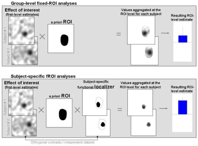
The standard method for dealing with this in group fMRI studies is to warp everyone's brain into a common space — Montreal Neurological Institute (MNI) space is most common — and then run statistics voxel by voxel. Where enough people show activation, you call it significant. This works for gross anatomy, but for high-level category-selective areas it loses resolution in two specific ways. Nieto-Castañón and Fedorenko (2012) showed mathematically that group analyses suffer from lower sensitivity (missing real effects when regions don't line up perfectly across subjects) and lower functional resolution (blurring together adjacent regions with different functions) 1.
The intuition is simple: if the FFA is 4 mm to the left in Subject A and 4 mm to the right in Subject B, a group average smears the signal across 8 mm of cortex that encompasses both the true FFA and some adjacent region doing something else entirely.
The fROI solution: localizers and independence
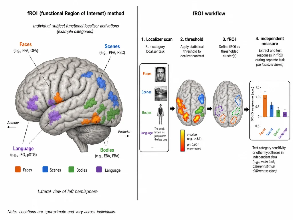
The fROI method bypasses group averaging entirely. The idea is to find each region in each person individually using a dedicated scan, and then test your scientific hypotheses against that person-specific region.
A typical experiment using fROIs has two phases:
Phase 1 — the localizer run. The subject views a blocked or event-related stimulus set designed to drive the region of interest. For the FFA, this means alternating blocks of faces and objects. For the PPA, it means scenes versus objects. For language regions in the frontal and temporal cortex, it means sentences versus meaningless word-strings. The localizer is designed to be a strong, unambiguous contrast that lights up the target region reliably.
Phase 2 — the experimental run. The subject performs the actual task of interest — viewing new stimuli, making judgments, remembering items, or whatever the hypothesis requires. These data are completely independent from the localizer.
The fROI is defined by thresholding the localizer contrast in each individual subject (typically at p < 0.001 uncorrected) and using the resulting cluster as a mask. You then measure the BOLD response within that subject's own mask during the experimental run.
This separation between the data that defines the region and the data that tests it is the critical safeguard. It eliminates "double dipping" — the circularity that would arise if you used the same data to find a region and then claim it responds to the thing you used to find it 2.
How Kanwisher invented it
Nancy Kanwisher describes the moment in her 2017 essay in Journal of Neuroscience. In 1995, her lab was trying to characterize the face-selective blob they'd found on the underside of the right hemisphere. They faced a statistics problem: they had found what looked like a real region, but any threshold correction for the tens of thousands of voxels in a whole-brain scan was either too liberal or too conservative 2.
The solution they invented was to split the data: use even-numbered runs to find the face-selective cluster in each subject, then extract the BOLD response from that cluster using only the odd-numbered runs. "Now I could run simple t-tests or ANOVAs across subjects on the resulting response magnitudes," Kanwisher writes. "No correction for multiple statistical comparisons was necessary because I was running a single statistical test on a single 'functional region of interest' or 'fROI.'"
What had looked like a statistics trick was actually a principled method for studying a thing in nature, not just a statistical blob. Once you have a localizer-defined fROI, you can ask open-ended questions about it: Does it respond more to photographs than cartoons? Does it respond when people imagine faces? Does it respond in blind individuals? Each new experiment uses the same localizer-defined region, so results can accumulate across labs and across years.
Kanwisher acknowledges the method wasn't entirely new — Roger Tootell had been mapping area MT and retinotopic areas V1–V5 with localizers since 1995 3, and single-unit neurophysiologists had always needed to identify what area their electrode was in before interpreting responses. But applying the approach systematically to high-level category-selective regions of human cortex, and building a cumulative research program around it, was what made the FFA paper's method as influential as its discovery.
The Group-Constrained Subject-Specific (GSS) method
The original fROI approach required researchers to eyeball each subject's activation map and manually select which cluster counted as the "true" FFA. This worked, but was subjective — two researchers looking at the same map might draw different boundaries.
Julian, Kanwisher and colleagues formalized the method in 2012 with the Group-Constrained Subject-Specific (GSS) algorithm 4. The logic has three steps:
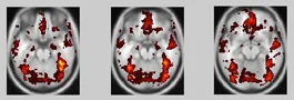
- Probabilistic overlap maps. Take each individual subject's thresholded localizer activation (faces > objects, p < 0.0001) and overlay all subjects in a common space. Each voxel gets a score: how many subjects activated there. This produces a heat map of where the FFA tends to be.
- Watershed parcels. Run a watershed image segmentation algorithm on the overlap map. This divides the map into "functional parcels" following its topology, like valleys separating hills. Only parcels where at least 60% of subjects show activation are retained. These parcels define candidate regions — they're essentially saying, "something consistent is happening here."
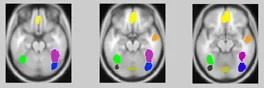
- Individual intersection. For each subject, intersect their own thresholded localizer activation with the pre-defined parcel outline. Whatever activated within the parcel boundary becomes that subject's personal fROI for that region.
The result is that the algorithm eliminates the subjective step while preserving the core logic. Every researcher who runs the GSS procedure on the same dataset will get the same parcels, and every subject's fROI is still located in their native anatomy rather than group-averaged coordinates. Julian et al. demonstrated the procedure for 14 well-studied category-selective areas in the ventral visual pathway — face areas, place areas, body areas, object areas.
Why this matters more than it sounds
The fROI method isn't just a statistical housekeeping trick. It changes what kind of science is possible.
Cumulative research. When every lab uses a localizer to define the FFA in each participant before measuring responses, they're all studying the same functional entity. Results replicate or fail to replicate against a consistent target, rather than against whatever cluster happened to survive threshold in a group analysis. Kanwisher calls this the ability to do "cognitive psychology on a little patch of the brain" — iterative hypothesis testing, experiment by experiment, converging on what the region actually represents.
Avoiding circular statistics. The "double dipping" problem identified by Kriegeskorte et al. (2009) was widespread in fMRI: researchers would find a region that responded to condition X, then extract the response to X from that region, and report an inflated effect size. The localizer-plus-independent-data structure rules this out structurally 2.
Cross-subject and cross-lab comparability. MNI coordinates fail at this for high-level regions because functional anatomy doesn't register to macroanatomy with enough precision. A reported "FFA peak at MNI [-42, -54, -18]" in one paper may not correspond well to the same peak in another lab's subjects. A localizer-defined fROI sidesteps this: you're always testing the face-selective region in this particular person, wherever it happens to sit.
What the method doesn't solve
The fROI approach has critics. Friston and colleagues (2006) argued that functional localizers are not neutral — the localizer contrast itself embeds theoretical assumptions about what the region does, and those assumptions constrain what you can discover with the resulting fROI 5. If you define the FFA as "the region that responds more to faces than to objects," you've built "face-selective" into the region's definition. The fROI can't then tell you whether the FFA is really face-selective — because you've defined it to be.
This is a genuine methodological tension. Kanwisher's response is that the experimental run is independent, so the localizer doesn't pre-bias the test results — only the choice of which region to study is affected. But if the true region is "the fusiform gyrus portion involved in expert object recognition," defining it with face > object will select the right cortical address while possibly missing some of its functional scope.
The fROI also doesn't resolve questions about what a region computes at a fine-grained level — MVPA and neural network modeling approaches are better suited to that. What fROIs do is carve out a stable object of study that can support decades of converging tests about function, connectivity, development, and causal role.
The broader program
The regions identified this way — FFA, PPA, OFA, EBA, the language network — now form a kind of map of functional architecture that Kanwisher has called "an initial sketch of the basic components of the human mind."
What's striking is how much that sketch has remained stable. Labs across the world, using different localizer designs, different scanner field strengths, and different participant populations, keep finding the same regions. The FFA shows up in the right fusiform gyrus. The PPA shows up in the parahippocampal gyrus. The EBA shows up in the lateral occipitotemporal cortex. The left frontal and temporal language areas activate for sentences more than non-sentences. This replicability is itself a result, not just a methodological nicety. It suggests that whatever these regions are doing, they're doing it consistently enough across human brains to be reliably localized with a short functional scan.
The same method that found the FFA in 1997 is still the workhorse of cognitive neuroscience today. The GSS algorithm made it fully automated; presurgical language mapping clinics use the same localizer logic to identify eloquent cortex in individual patients before tumor resection 6. The fROI isn't just a research tool — it's now the standard for understanding what any individual brain is doing.
Landmark paper: Julian, J.B., Fedorenko, E., Webster, J., & Kanwisher, N. (2012). An algorithmic method for functionally defining regions of interest in the ventral visual pathway. NeuroImage, 60(4), 2357–2364. 4
Course connection: MIT 9.13 The Human Brain — "Why Use Functional Regions of Interest (fROIs)?" video lecture by Nancy Kanwisher: nancysbraintalks.mit.edu
Fuentes de referencia
- 1Subject-specific functional localizers increase sensitivity and functional resolution — Nieto-Castañón & Fedorenko 2012
- 2The Quest for the FFA and Where It Led — Kanwisher 2017, Journal of Neuroscience
- 3Sereno et al. 1995, retinotopy — Science
- 4An algorithmic method for functionally defining regions of interest — Julian et al. 2012
- 5A critique of functional localisers — Friston et al. 2006
- 6Subject-specific fROIs enhance reliability in presurgical diagnostics — Van Kube et al. 2025
Añade más opiniones o contexto en torno a este contenido.